Copyright
©2011 Baishideng Publishing Group Co.
World J Gastrointest Endosc. May 16, 2011; 3(5): 101-104
Published online May 16, 2011. doi: 10.4253/wjge.v3.i5.101
Published online May 16, 2011. doi: 10.4253/wjge.v3.i5.101
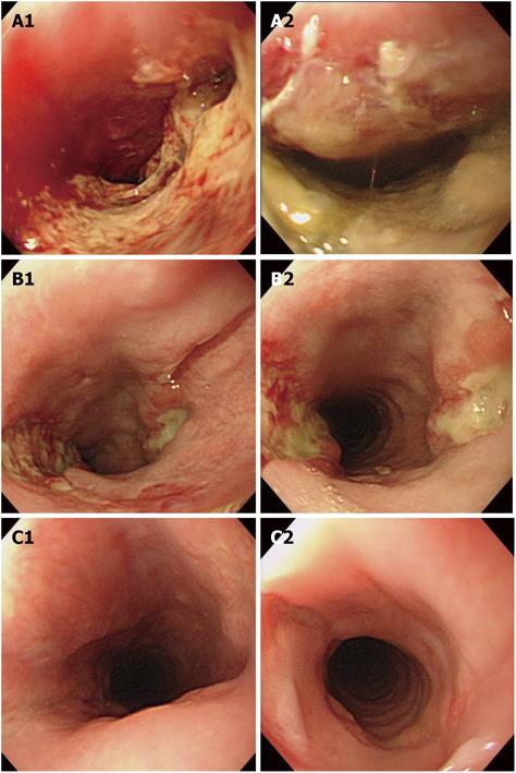
Figure 1 Course of endoscopic findings.
A: Findings on initial endoscopy (day 3). An ulcerated lesion covered by a thick white coating is seen in the upper esophagus. The esophageal lumen is narrowed, with easy friability. No distinct surrounding ridge is evident, but ulcer margins are irregular; B: Findings on second endoscopy (day 9). Deep ulceration with a whitish coating is seen at the 3 and 9 o’clock positions in the lumen. Compared to the initial endoscopic findings, esophageal stenosis has improved; C: Findings on third endoscopy (day 23). The white coating has disappeared, and the esophageal ulcer is healed and scarred.
- Citation: Enomoto S, Nakazawa K, Ueda K, Mori Y, Maeda Y, Shingaki N, Maekita T, Ota U, Oka M, Ichinose M. Steakhouse syndrome causing large esophageal ulcer and stenosis. World J Gastrointest Endosc 2011; 3(5): 101-104
- URL: https://www.wjgnet.com/1948-5190/full/v3/i5/101.htm
- DOI: https://dx.doi.org/10.4253/wjge.v3.i5.101


