Copyright
©2011 Baishideng Publishing Group Co.
World J Gastrointest Endosc. Nov 16, 2011; 3(11): 209-212
Published online Nov 16, 2011. doi: 10.4253/wjge.v3.i11.209
Published online Nov 16, 2011. doi: 10.4253/wjge.v3.i11.209
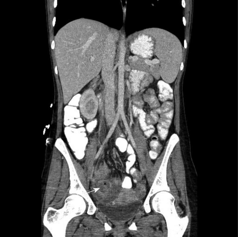
Figure 1 A 33-year-old female patient with a 3 cm pelvic abscess due to Crohn’s disease.
The computed tomography demonstrates an abscess cavity with a small air bubble within it (arrow) prior to drainage, which was accomplished by placing a drainage catheter via the transgluteal route.
- Citation: Richards RJ. Management of abdominal and pelvic abscess in Crohn’s disease. World J Gastrointest Endosc 2011; 3(11): 209-212
- URL: https://www.wjgnet.com/1948-5190/full/v3/i11/209.htm
- DOI: https://dx.doi.org/10.4253/wjge.v3.i11.209


