Copyright
©2011 Baishideng Publishing Group Co.
World J Gastrointestinal Endoscopy. Oct 16, 2011; 3(10): 183-194
Published online Oct 16, 2011. doi: 10.4253/wjge.v3.i10.183
Published online Oct 16, 2011. doi: 10.4253/wjge.v3.i10.183
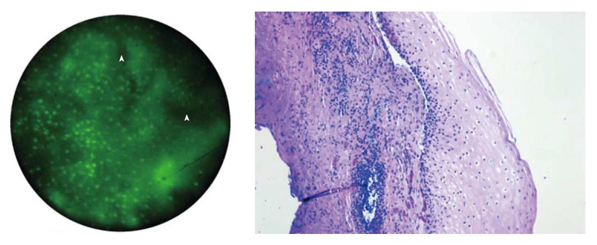
Figure 2 Images of normal squamous tissue using the High Resolution Microendoscope.
A: Endoscopic microscope image of normal squamous tissue stained with 0.05% acriflavine shows flat arrangement of squamous epithelium with round regularly spaced nuclei. The round clear spaces surrounded by the epithelium represent the papillae (arrowhead). The acriflavine in image A highlights the nuclei; B: Histopathology of same specimen. Scale bar is 100 microns.
- Citation: Shukla R, Abidi WM, Richards-Kortum R, Anandasabapathy S. Endoscopic imaging: How far are we from real-time histology? World J Gastrointestinal Endoscopy 2011; 3(10): 183-194
- URL: https://www.wjgnet.com/1948-5190/full/v3/i10/183.htm
- DOI: https://dx.doi.org/10.4253/wjge.v3.i10.183


