Copyright
©2011 Baishideng Publishing Group Co.
World J Gastrointest Endosc. Jan 16, 2011; 3(1): 11-15
Published online Jan 16, 2011. doi: 10.4253/wjge.v3.i1.11
Published online Jan 16, 2011. doi: 10.4253/wjge.v3.i1.11
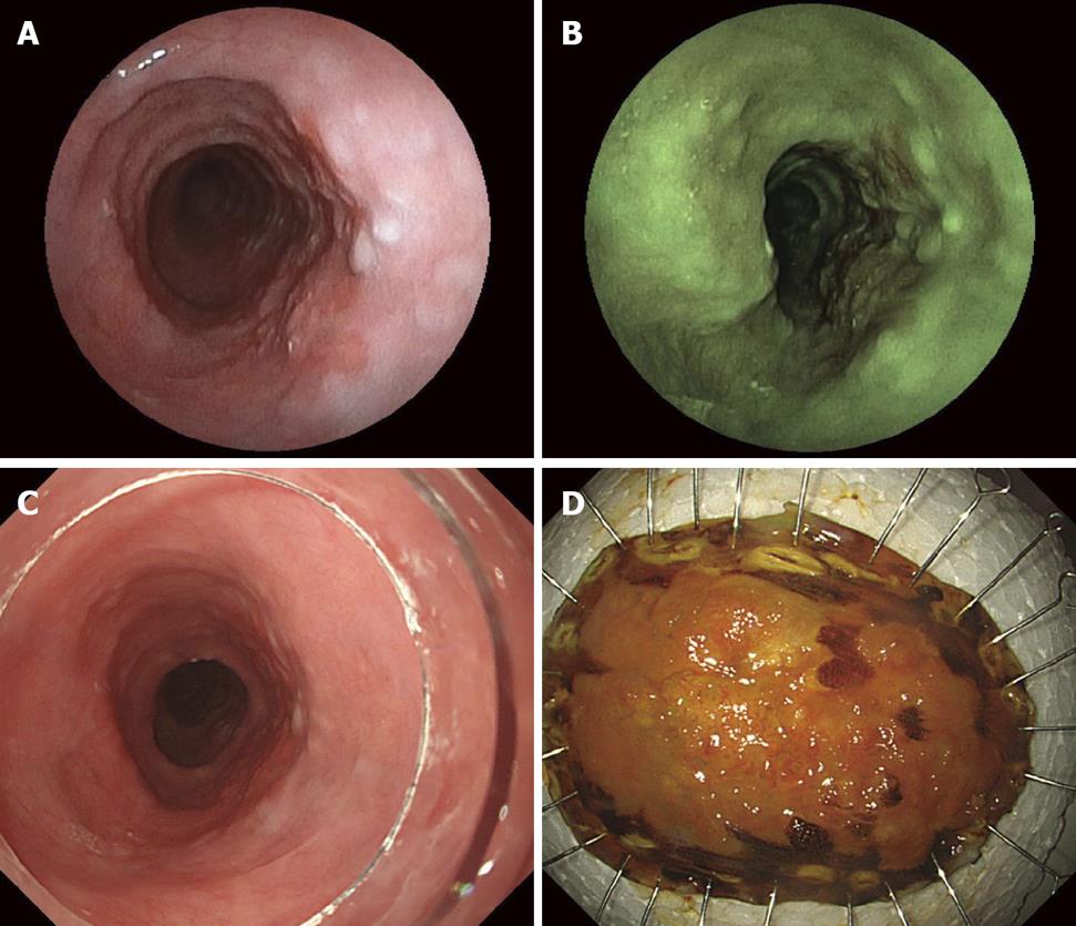
Figure 3 A rough, slightly elevated squamous lesion with hyperemia in a semicircle is found 25 cm distal from the superior incisor line.
The hyperemia of the lesion is highlighted and the boundary is more clearly visualized with flexible spectral imaging color enwhancement (FICE). The visibility was graded 4. Endoscopic submucosal dissection (ESD) was performed for local complete resection of the squamous cell carcinoma including the muscularis mucosae. A: Conventional image with ultraslim endoscopy: A rough, elevated squamous lesion with hyperemia in a semicircle is found; B: Image with FICE: The hyperemia of the lesion is highlighted and the boundary is more clearly visualized with FICE (FICE 4); C: Conventional image with conventional esophagogastroduodenoscopy; D: Dissected section sprayed with Iodin after ESD.
- Citation: Tanioka Y, Yanai H, Sakaguchi E. Ultraslim endoscopy with flexible spectral imaging color enhancement for upper gastrointestinal neoplasms. World J Gastrointest Endosc 2011; 3(1): 11-15
- URL: https://www.wjgnet.com/1948-5190/full/v3/i1/11.htm
- DOI: https://dx.doi.org/10.4253/wjge.v3.i1.11


