Copyright
©2011 Baishideng Publishing Group Co.
World J Gastrointest Endosc. Jan 16, 2011; 3(1): 11-15
Published online Jan 16, 2011. doi: 10.4253/wjge.v3.i1.11
Published online Jan 16, 2011. doi: 10.4253/wjge.v3.i1.11
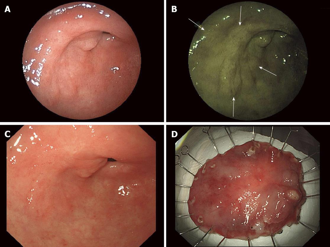
Figure 2 A discolored change was found in the anterior wall and surrounding areas of the gastric antrum.
Color contrast between the lesion and surrounding mucosa with atrophic changes is highlighted and the boundary is more clearly visualized with flexible spectral imaging color enhancement (FICE). The visibility with FICE was graded 4. Endoscopic submucosal dissection (ESD) was performed for local complete resection of the well-differentiated adenocarcinoma limited to within the mucosa. A: Conventional image with ultraslim endoscopy; B: Image with FICE: Color contrast between the lesion and surrounding mucosa is highlighted and the boundary is more clearly visualized (FICE 4); C: Conventional image with conventional esophagogastroduodenoscopy; D: Dissected section after ESD.
- Citation: Tanioka Y, Yanai H, Sakaguchi E. Ultraslim endoscopy with flexible spectral imaging color enhancement for upper gastrointestinal neoplasms. World J Gastrointest Endosc 2011; 3(1): 11-15
- URL: https://www.wjgnet.com/1948-5190/full/v3/i1/11.htm
- DOI: https://dx.doi.org/10.4253/wjge.v3.i1.11


