Copyright
©2011 Baishideng Publishing Group Co.
World J Gastrointest Endosc. Jan 16, 2011; 3(1): 1-5
Published online Jan 16, 2011. doi: 10.4253/wjge.v3.i1.1
Published online Jan 16, 2011. doi: 10.4253/wjge.v3.i1.1
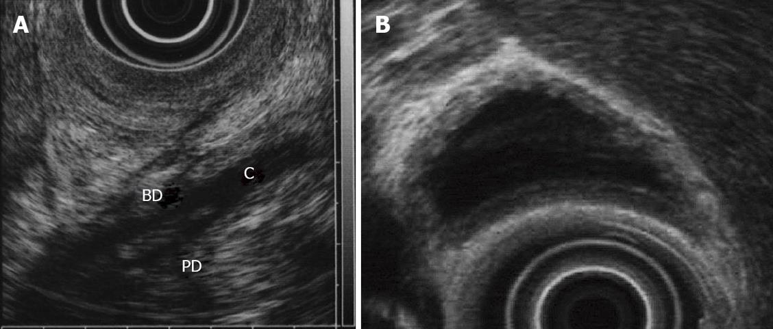
Figure 5 Endoscopic ultrasonography of a pancreaticobiliary maljunction patient.
A:The confluence of pancreatic duct and bile duct in the proximal portion of the duodenal wall; B: thickness of inner low echoic layer of the gallbladder. BD: bile duct; PD: pancreatic duct; C: common channel.
- Citation: Kamisawa T, Takuma K, Itokawa F, Itoi T. Endoscopic diagnosis of pancreaticobiliary maljunction. World J Gastrointest Endosc 2011; 3(1): 1-5
- URL: https://www.wjgnet.com/1948-5190/full/v3/i1/1.htm
- DOI: https://dx.doi.org/10.4253/wjge.v3.i1.1


