Copyright
©2010 Baishideng.
World J Gastrointest Endosc. Aug 16, 2010; 2(8): 288-292
Published online Aug 16, 2010. doi: 10.4253/wjge.v2.i8.288
Published online Aug 16, 2010. doi: 10.4253/wjge.v2.i8.288
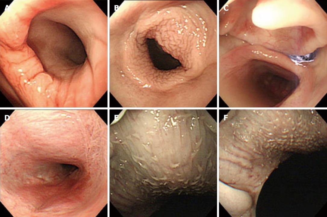
Figure 2 Images of retrograde observation.
A: Esophagogastric junction, and the entire view is provided in a single visual field; B: Observation of the hypopharyngoesophageal junction from the cervical esophagus. The cervical esophagus is dilated well, providing a good visual field; C: Laryngeal surface of the epiglottis viewed up from the hypopharynx. Epipharynx and nasal cavity are present in the arrow direction; D: Observation of the epipharynx is possible; E, F: Observation of the lingual root is possible.
- Citation: Honda M, Hori Y, Shionoya Y, Nakada A, Sato T, Kobayashi T, Shimada H, Kida N, Nakamura T. Observation of the esophagus, pharynx and lingual root by gastrointestinal endoscopy with a percutaneous retrograde approach. World J Gastrointest Endosc 2010; 2(8): 288-292
- URL: https://www.wjgnet.com/1948-5190/full/v2/i8/288.htm
- DOI: https://dx.doi.org/10.4253/wjge.v2.i8.288


