Copyright
©2010 Baishideng.
World J Gastrointest Endosc. Aug 16, 2010; 2(8): 278-287
Published online Aug 16, 2010. doi: 10.4253/wjge.v2.i8.278
Published online Aug 16, 2010. doi: 10.4253/wjge.v2.i8.278
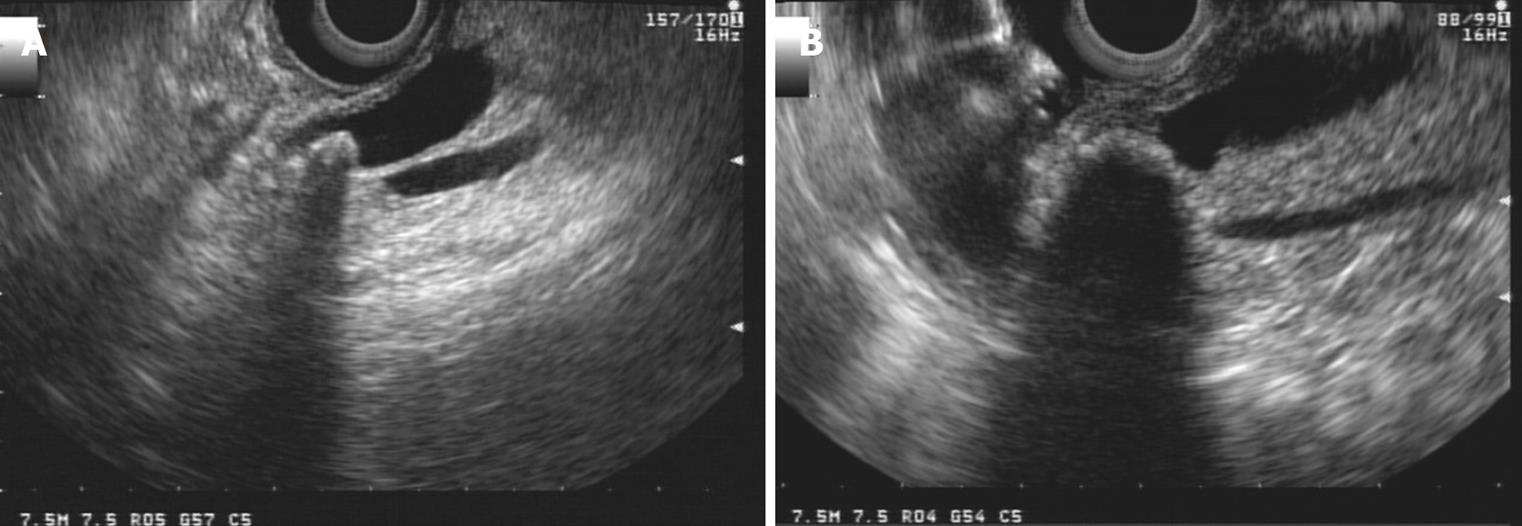
Figure 12 Lineal imaging of distal choledochal lithiasis.
A: Stone and acoustic shadow is clearly seen; B: Bulging of the papilla due to impacted stone.
- Citation: Castillo C. Endoscopic ultrasound in the papilla and the periampullary region. World J Gastrointest Endosc 2010; 2(8): 278-287
- URL: https://www.wjgnet.com/1948-5190/full/v2/i8/278.htm
- DOI: https://dx.doi.org/10.4253/wjge.v2.i8.278


