Copyright
©2010 Baishideng.
World J Gastrointest Endosc. Aug 16, 2010; 2(8): 271-277
Published online Aug 16, 2010. doi: 10.4253/wjge.v2.i8.271
Published online Aug 16, 2010. doi: 10.4253/wjge.v2.i8.271
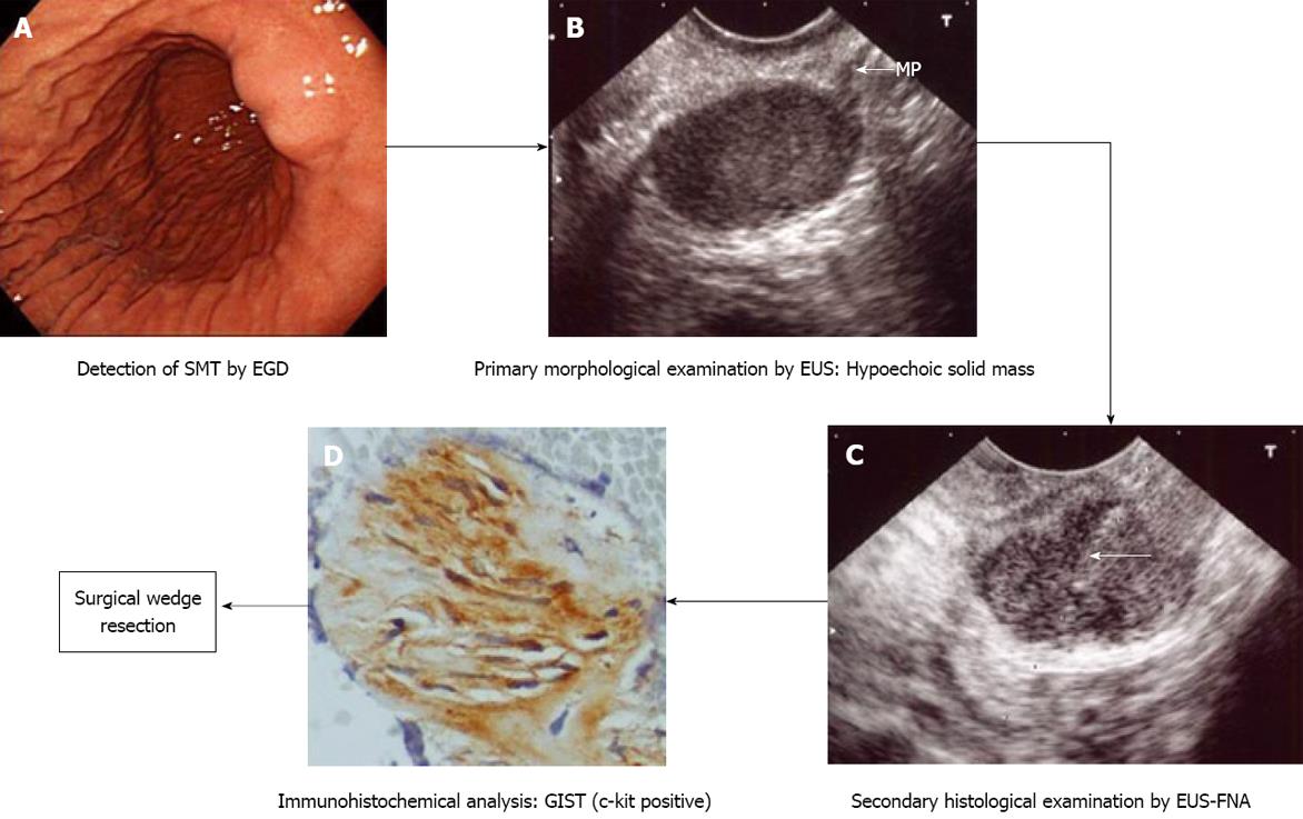
Figure 3 Management process of gastric submucosal tumor (in a case of gastrointestinal stromal tumor) according to our institutional algorithm (Figure 1).
Quoted and modified from reference [15]. A: Esophagogastroduodenoscopy (EGD) shows submucosal tumor in the lower body of the stomach; B: Endoscopic ultrasound (EUS) reveals 2.5 cm subepithelial hypoechoic solid tumor with continuity to proper muscle layer (arrow-mp); C: Puncture of the small gastrointestinal stromal tumor (GIST) under EUS guidance. Arrow: tip of needle; D: The immunohistochemical fnding of endoscopic ultrasound-guided fne needle aspiration (EUS-FNA) specimen of GIST. The tumor is diffusely positive for c-kit.
- Citation: Akahoshi K, Oya M. Gastrointestinal stromal tumor of the stomach: How to manage? World J Gastrointest Endosc 2010; 2(8): 271-277
- URL: https://www.wjgnet.com/1948-5190/full/v2/i8/271.htm
- DOI: https://dx.doi.org/10.4253/wjge.v2.i8.271


