Copyright
©2010 Baishideng.
World J Gastrointest Endosc. Mar 16, 2010; 2(3): 90-96
Published online Mar 16, 2010. doi: 10.4253/wjge.v2.i3.90
Published online Mar 16, 2010. doi: 10.4253/wjge.v2.i3.90
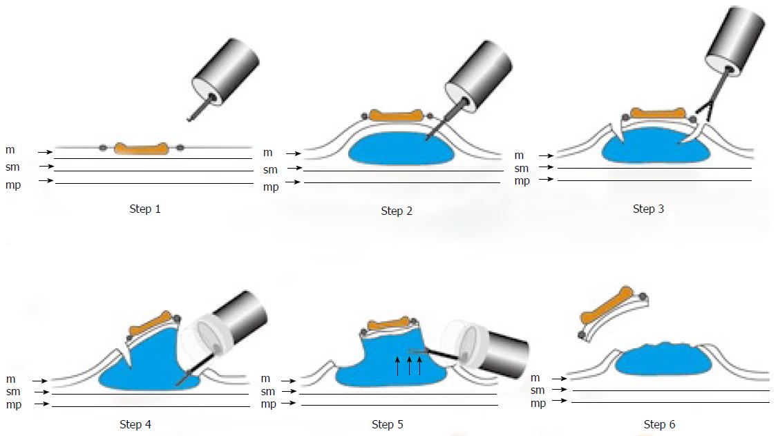
Figure 3 Schematic shows ESD using GSF.
Step 1: Marking dots are made on the circumference of the lesion to outline the incision line; Step 2: A concentrated glycerin solution mixed with a small volume of epinephrine and indigo carmine dye is injected into the submucosal layer around the target lesion to lift the entire lesion; Step 3: The lesion is separated from the surrounding normal mucosa by complete incision around the lesion using the GSF; Step 4: A piece of submucosal tissue is grasped by GSF; Step5: A grasped tissue is lifted up and cut with the GSF using electrosurgical current to effect submucosal exfoliation; Step 6: The lesion is resected in one piece; m: Mucosa; sm: Submucosa; mp: Muscularis propria.
- Citation: Akahoshi K, Akahane H. A new breakthrough: ESD using a newly developed grasping type scissor forceps for early gastrointestinal tract neoplasms. World J Gastrointest Endosc 2010; 2(3): 90-96
- URL: https://www.wjgnet.com/1948-5190/full/v2/i3/90.htm
- DOI: https://dx.doi.org/10.4253/wjge.v2.i3.90


