Copyright
©The Author(s) 2025.
World J Gastrointest Endosc. Apr 16, 2025; 17(4): 105238
Published online Apr 16, 2025. doi: 10.4253/wjge.v17.i4.105238
Published online Apr 16, 2025. doi: 10.4253/wjge.v17.i4.105238
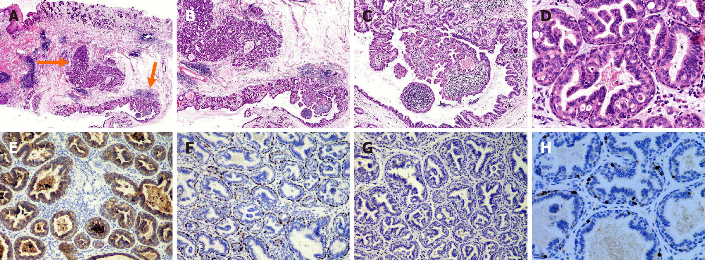
Figure 2 Pathological images of esophageal submucosal gland duct adenoma.
A: The orange arrow denoted the submucosal lesion within the esophagus (original magnification × 40); B and C: The lesion was composed of ducts or cysts containing papillary and tubular structures with lymphocytic infiltrates in the interstitial (B: Original magnification × 100, C: Original magnification × 200); D: Tumor cells were composed of moderate bilayer epithelium with no mitotic figures (original magnification × 400); E: CK7 was positive in inner cells and negative in outer cells (original magnification × 200); F: P63 was negative in inner cells and positive in outer cells (original magnification × 200); G: All cells were negative for MUC5AC (original magnification × 200); H: Ki-67 showed only expression in a few myoepithelial cells(original magnification × 400).
- Citation: Lu T, Liu JX, Xia Y, Zhao Y. Clinical, endoscopic and histopathological observation of a rare case of esophageal submucosal gland duct adenoma: A case report. World J Gastrointest Endosc 2025; 17(4): 105238
- URL: https://www.wjgnet.com/1948-5190/full/v17/i4/105238.htm
- DOI: https://dx.doi.org/10.4253/wjge.v17.i4.105238


