Copyright
©The Author(s) 2025.
World J Gastrointest Endosc. Feb 16, 2025; 17(2): 102075
Published online Feb 16, 2025. doi: 10.4253/wjge.v17.i2.102075
Published online Feb 16, 2025. doi: 10.4253/wjge.v17.i2.102075
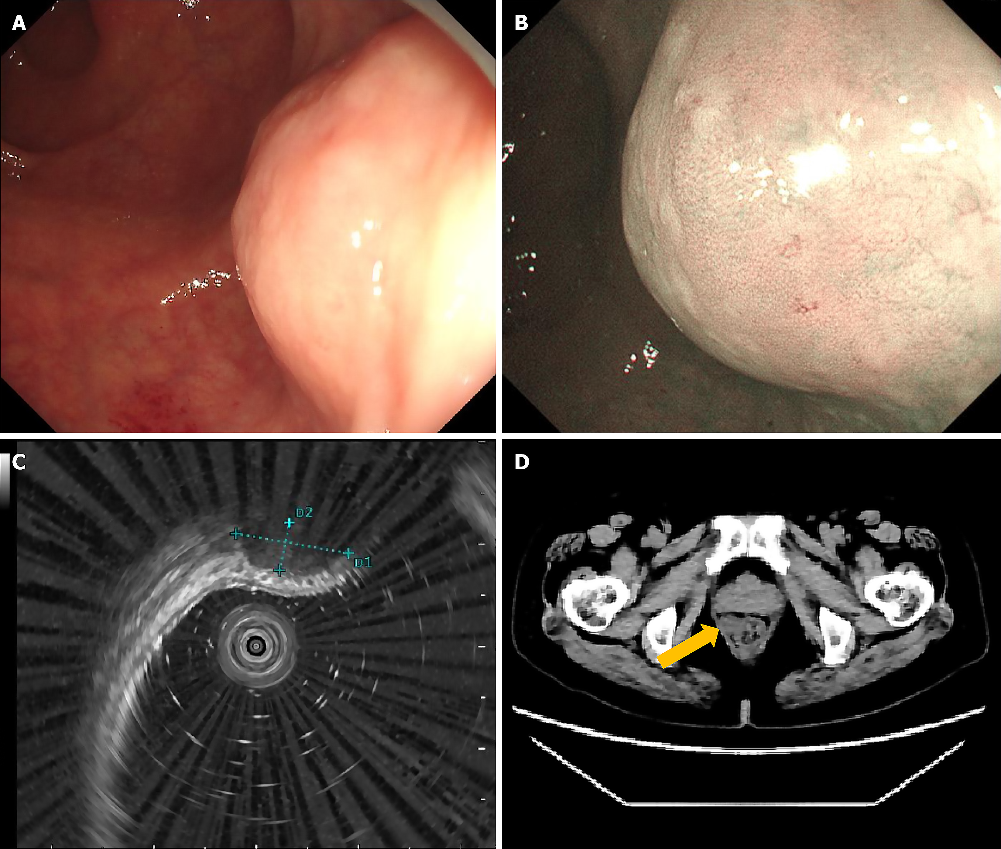
Figure 1 Colonoscopy and abdominopelvic computed tomography.
A: White light endoscopy; B: Narrow band imaging; C: Endoscopic ultrasonography (a small probe at 20 Hz) showing a hypoechoic mass originating from the muscularis propria with clear boundary; D: A well-circumscribed, well-enhanced, round-shaped mass was identified at the rectum; the main growth pattern was extraluminal growth (yellow arrow).
- Citation: Zhang YJ, Yuan MX, Wen W, Jian Y, Zhang CM, Yuan J, He L. Endoscopic full-thickness resection of rectal schwannoma: A case report. World J Gastrointest Endosc 2025; 17(2): 102075
- URL: https://www.wjgnet.com/1948-5190/full/v17/i2/102075.htm
- DOI: https://dx.doi.org/10.4253/wjge.v17.i2.102075


