Copyright
©The Author(s) 2025.
World J Gastrointest Endosc. Feb 16, 2025; 17(2): 101135
Published online Feb 16, 2025. doi: 10.4253/wjge.v17.i2.101135
Published online Feb 16, 2025. doi: 10.4253/wjge.v17.i2.101135
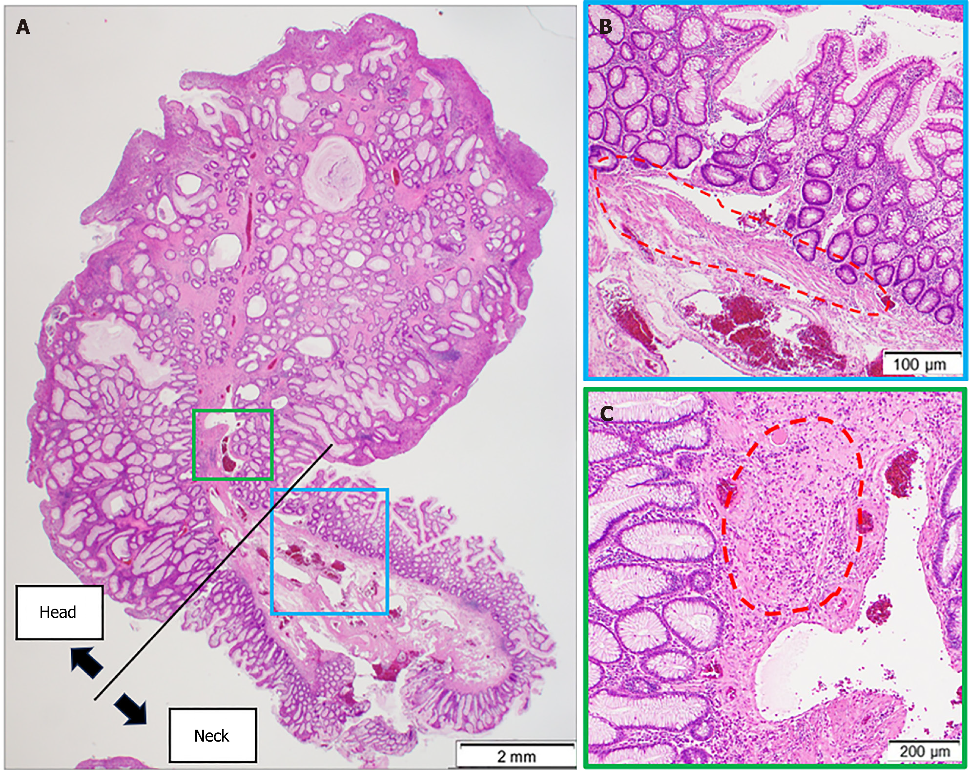
Figure 5 Histopathological evaluation of juvenile polyps.
A: Glandular ducts with dilated lumens distributed in the polyp; B: At the stem of the polyp (blue box in panel A, the lamina muscularis mucosae are present (dotted circle); C: Whereas in the head (green box in panel A), the lamina muscularis mucosae are lacking and a high inflammatory cell infiltrate is present (dotted circle).
- Citation: Kataoka F, Nakanishi T, Araki H, Ichino S, Kamei M, Makino H, Nagao R, Asano T, Tagami A, Moriwaki H. Adult juvenile polyp bleeding detected by extravascular contrast leakage and treated with endoscopic clipping: A case report. World J Gastrointest Endosc 2025; 17(2): 101135
- URL: https://www.wjgnet.com/1948-5190/full/v17/i2/101135.htm
- DOI: https://dx.doi.org/10.4253/wjge.v17.i2.101135


