Copyright
©The Author(s) 2024.
World J Gastrointest Endosc. Sep 16, 2024; 16(9): 533-539
Published online Sep 16, 2024. doi: 10.4253/wjge.v16.i9.533
Published online Sep 16, 2024. doi: 10.4253/wjge.v16.i9.533
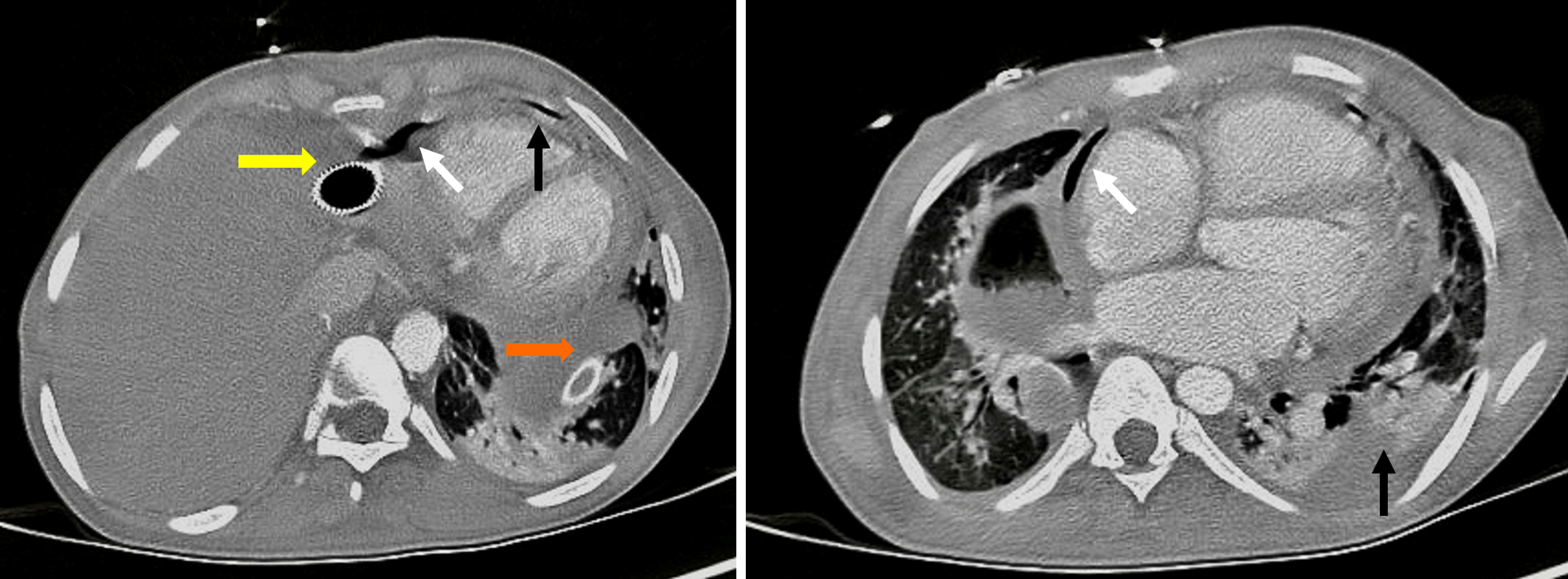
Figure 1 Axial chest tomography with contrast demonstrating a fistulous tract indicated by air density in the pericardium (white arrows) and the left pleural space with complex effusion (black arrows).
Note the self-expandable metallic esophageal prosthesis (orange arrow) and the thoracostomy tube (yellow arrow).
- Citation: Muñoz-González S, Quejada-Cuesta S, González-Arroyave D, Ardila CM. Endoscopic vacuum assisted closure therapy for esophagopericardial fistula in a 16-year-old male: A case report. World J Gastrointest Endosc 2024; 16(9): 533-539
- URL: https://www.wjgnet.com/1948-5190/full/v16/i9/533.htm
- DOI: https://dx.doi.org/10.4253/wjge.v16.i9.533


