Copyright
©The Author(s) 2024.
World J Gastrointest Endosc. Aug 16, 2024; 16(8): 494-499
Published online Aug 16, 2024. doi: 10.4253/wjge.v16.i8.494
Published online Aug 16, 2024. doi: 10.4253/wjge.v16.i8.494
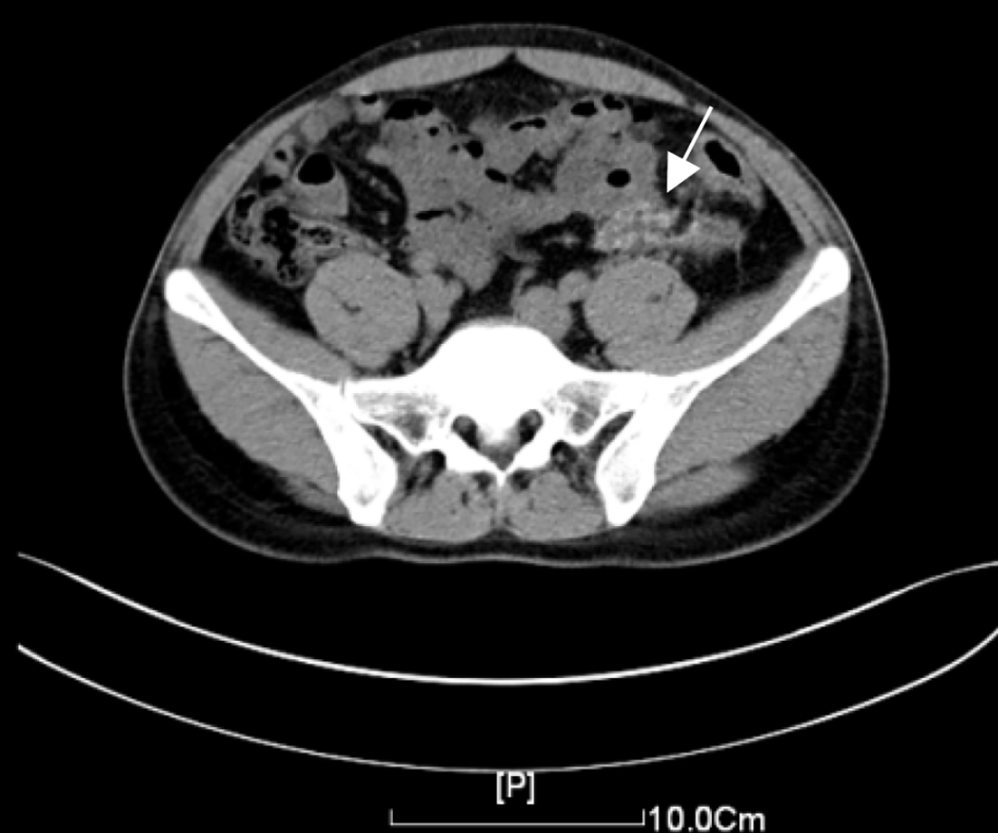
Figure 1 Computed tomography scan.
Image revealed irregular thickening of the left descending colon wall, luminal narrowing, an irregular patchy infiltrative density in the surrounding mesentery, and internal calcifications, measuring approximately 3.40 cm × 1.70 cm in cross-sectional size.
- Citation: Zhang BF, Liu J, Zhang S, Chen L, Lu JZ, Zhang MQ. Heterotopic mesenteric ossification caused by trauma: A case report. World J Gastrointest Endosc 2024; 16(8): 494-499
- URL: https://www.wjgnet.com/1948-5190/full/v16/i8/494.htm
- DOI: https://dx.doi.org/10.4253/wjge.v16.i8.494


