Copyright
©The Author(s) 2024.
World J Gastrointest Endosc. Aug 16, 2024; 16(8): 489-493
Published online Aug 16, 2024. doi: 10.4253/wjge.v16.i8.489
Published online Aug 16, 2024. doi: 10.4253/wjge.v16.i8.489
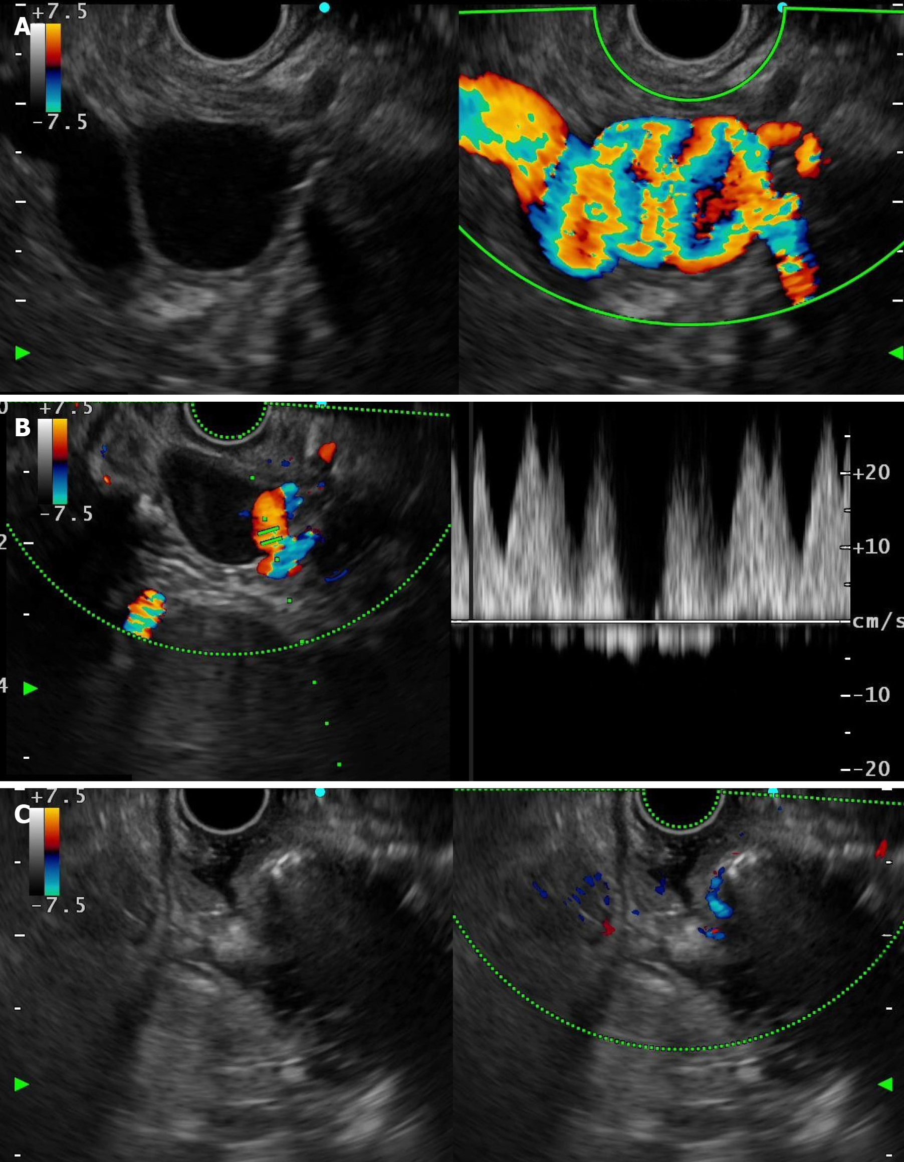
Figure 2 Endoscopic ultrasound-guided therapy.
A: Endoscopic ultrasonography shows varicose veins with a maximum diameter of 2.0 cm; B: Endoscopic ultrasonography shows a blood vessel entwined with varicose veins, and colour Doppler shows a pulsatile signal; C: Endoscopic ultrasound-guided coil embolization combined with cyanoacrylate injection.
- Citation: Zhang HY, He CC, Zhong DF. Endoscopic ultrasound-guided treatment of isolated gastric varices entwined with arteries: A case report. World J Gastrointest Endosc 2024; 16(8): 489-493
- URL: https://www.wjgnet.com/1948-5190/full/v16/i8/489.htm
- DOI: https://dx.doi.org/10.4253/wjge.v16.i8.489


