Copyright
©The Author(s) 2024.
World J Gastrointest Endosc. Jul 16, 2024; 16(7): 432-438
Published online Jul 16, 2024. doi: 10.4253/wjge.v16.i7.432
Published online Jul 16, 2024. doi: 10.4253/wjge.v16.i7.432
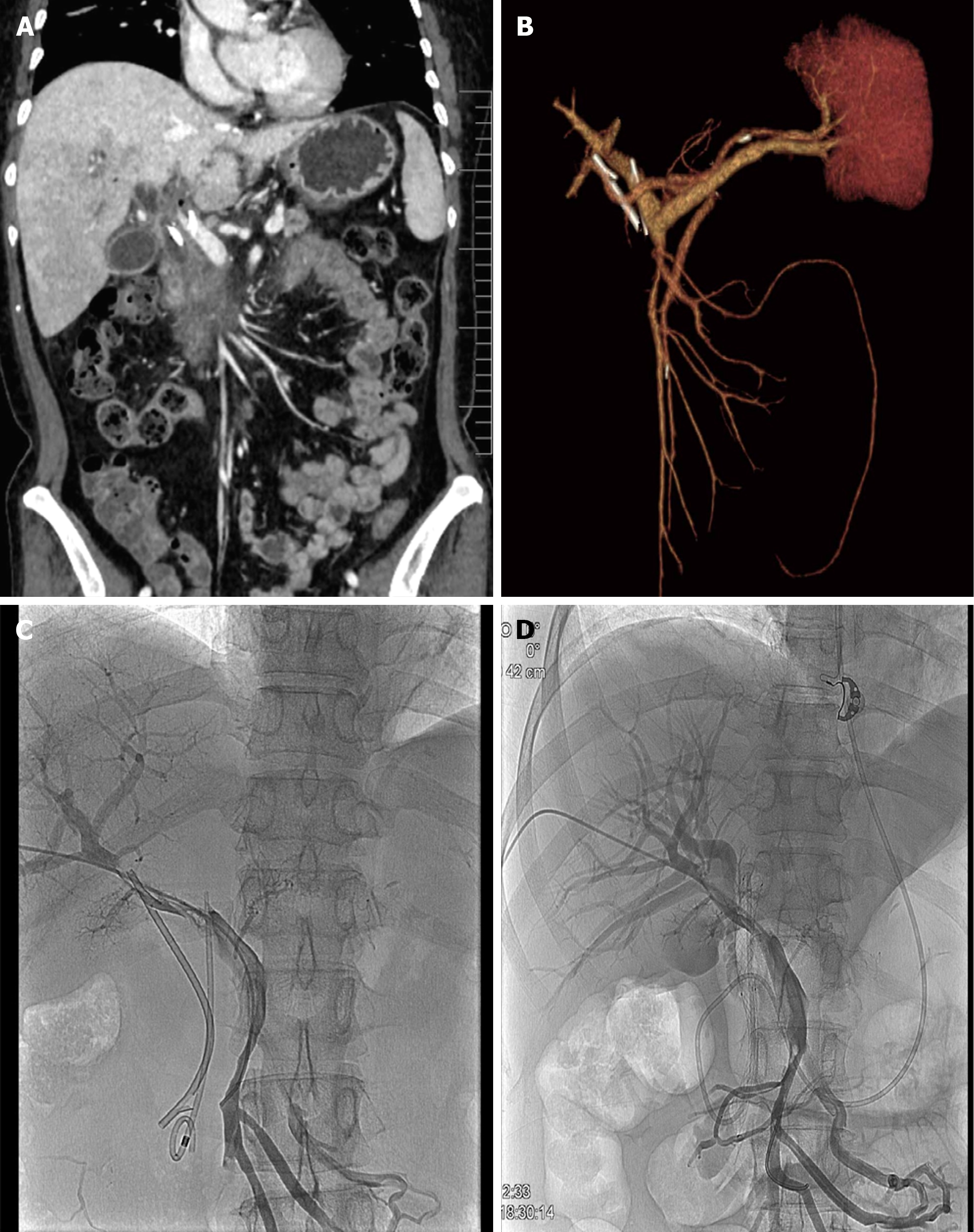
Figure 2 Contrast-enhanced computed tomography.
A: Contrast-enhanced computed tomography (CT) image showing that a biliary stent had entered the main portal vein; B: Contrast-enhanced CT images of the reconstructed portal vein; C: The single pigtail stent separated from the single flap stent and migrated straight up the spine into the portal vein; D: Repeated portal angiography showed no contrast leakage after placement of the fully covered metal stent in the common bile duct.
- Citation: Wu R, Zhang F, Zhu H, Liu MD, Zhuge YZ, Wang L, Zhang B. Recognition and management of stent malposition in the portal vein during endoscopic retrograde cholangiopancreatography: A case report. World J Gastrointest Endosc 2024; 16(7): 432-438
- URL: https://www.wjgnet.com/1948-5190/full/v16/i7/432.htm
- DOI: https://dx.doi.org/10.4253/wjge.v16.i7.432


