Copyright
©The Author(s) 2024.
World J Gastrointest Endosc. Jun 16, 2024; 16(6): 368-375
Published online Jun 16, 2024. doi: 10.4253/wjge.v16.i6.368
Published online Jun 16, 2024. doi: 10.4253/wjge.v16.i6.368
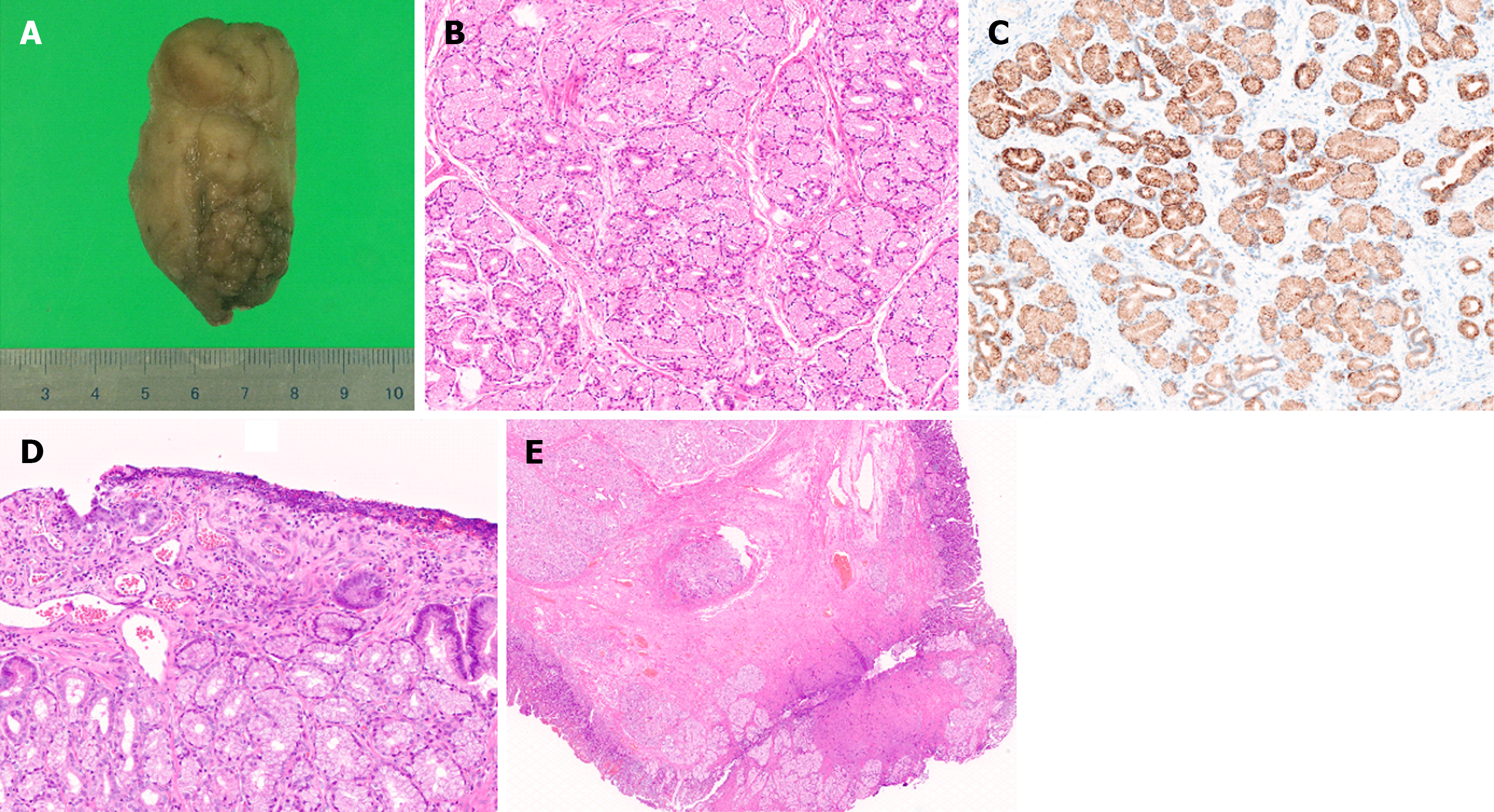
Figure 6 Pathological findings of the lesion.
A: The lesion size after formalin fixation was 6 cm × 3.5 cm × 3 cm; B: Histopathological examination revealed nodular proliferation of Brunner's glands without atypia, partitioned by fibrous septa; C: This site exhibited positive staining for MUC6; D: The area observed as a depressed region during EGD showed superficial erosion and regenerating epithelium; E: At the resection margin, thickened muscularis mucosa, fibrosis in the submucosal layer, and normal Brunner's glands were observed.
- Citation: Makazu M, Sasaki A, Ichita C, Sumida C, Nishino T, Nagayama M, Teshima S. Giant Brunner's gland hyperplasia of the duodenum successfully resected en bloc by endoscopic mucosal resection: A case report. World J Gastrointest Endosc 2024; 16(6): 368-375
- URL: https://www.wjgnet.com/1948-5190/full/v16/i6/368.htm
- DOI: https://dx.doi.org/10.4253/wjge.v16.i6.368


