Copyright
©The Author(s) 2024.
World J Gastrointest Endosc. Jun 16, 2024; 16(6): 368-375
Published online Jun 16, 2024. doi: 10.4253/wjge.v16.i6.368
Published online Jun 16, 2024. doi: 10.4253/wjge.v16.i6.368
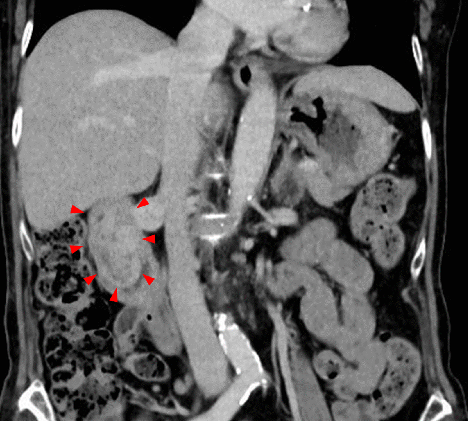
Figure 4 Abdominal contrast-enhanced computed tomography.
It revealed a 7 cm protruding lesion (arrowhead) hanging from the bulb to the descending portion of the duodenum. The lesion showed minimal enhancement in the arterial phase and a mosaic-like enhancement pattern in the venous phase.
- Citation: Makazu M, Sasaki A, Ichita C, Sumida C, Nishino T, Nagayama M, Teshima S. Giant Brunner's gland hyperplasia of the duodenum successfully resected en bloc by endoscopic mucosal resection: A case report. World J Gastrointest Endosc 2024; 16(6): 368-375
- URL: https://www.wjgnet.com/1948-5190/full/v16/i6/368.htm
- DOI: https://dx.doi.org/10.4253/wjge.v16.i6.368


