Copyright
©The Author(s) 2024.
World J Gastrointest Endosc. Mar 16, 2024; 16(3): 157-167
Published online Mar 16, 2024. doi: 10.4253/wjge.v16.i3.157
Published online Mar 16, 2024. doi: 10.4253/wjge.v16.i3.157
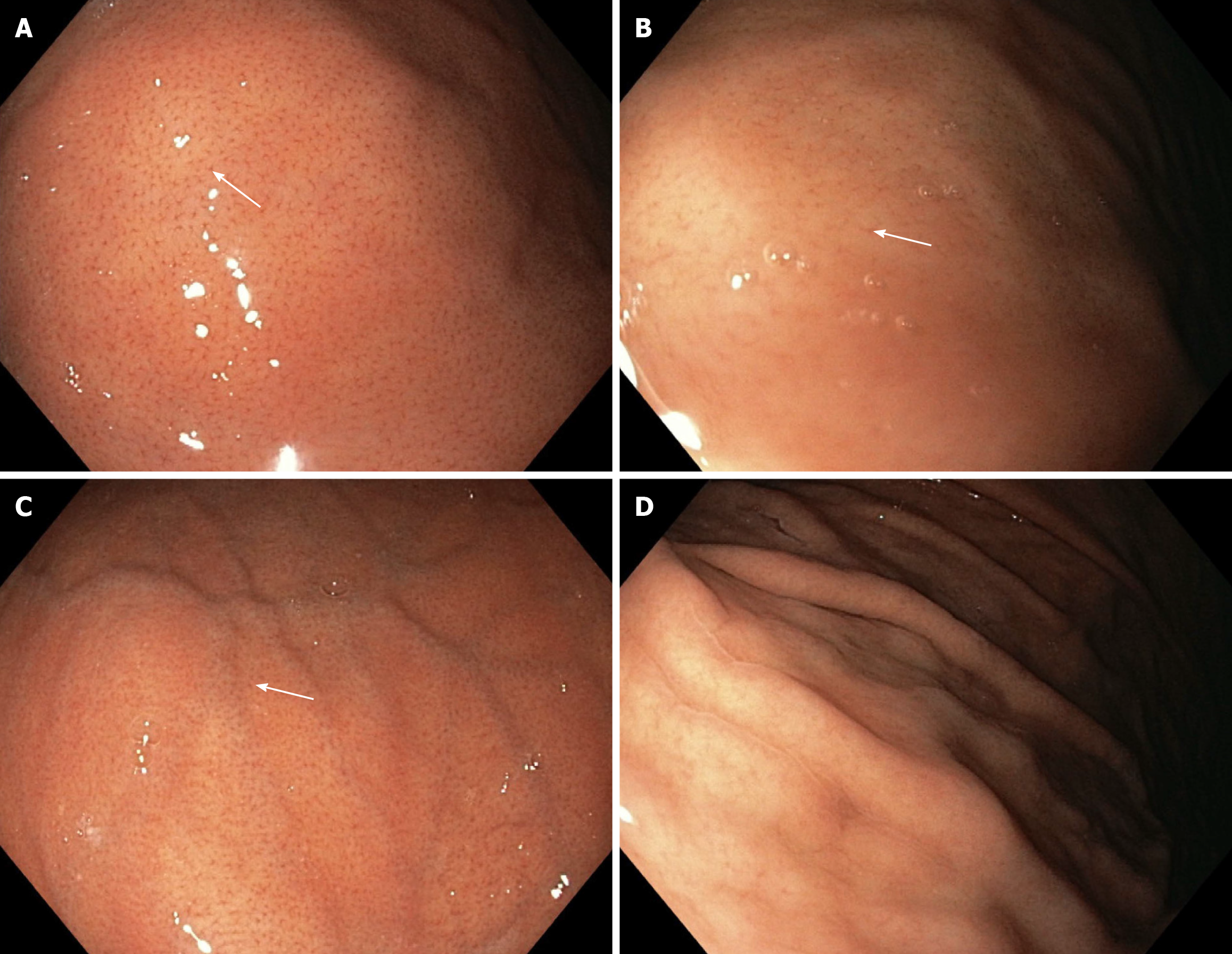
Figure 1 White light endoscopy from the point of view of the first observer.
A and B: White light endoscopy (WLE)-1a pattern and WLE-2a pattern shows a clear appearance of the regular arrangement of collecting venules (RAC) (white arrow); C: WLE-b pattern shows a less clear appearance of the RAC (white arrow); D: WLE-c pattern shows the absence of the RAC.
- Citation: Kurtcehajic A, Zerem E, Bokun T, Alibegovic E, Kunosic S, Hujdurovic A, Tursunovic A, Ljuca K. Could near focus endoscopy, narrow-band imaging, and acetic acid improve the visualization of microscopic features of stomach mucosa? World J Gastrointest Endosc 2024; 16(3): 157-167
- URL: https://www.wjgnet.com/1948-5190/full/v16/i3/157.htm
- DOI: https://dx.doi.org/10.4253/wjge.v16.i3.157


