Copyright
©The Author(s) 2024.
World J Gastrointest Endosc. Dec 16, 2024; 16(12): 678-685
Published online Dec 16, 2024. doi: 10.4253/wjge.v16.i12.678
Published online Dec 16, 2024. doi: 10.4253/wjge.v16.i12.678
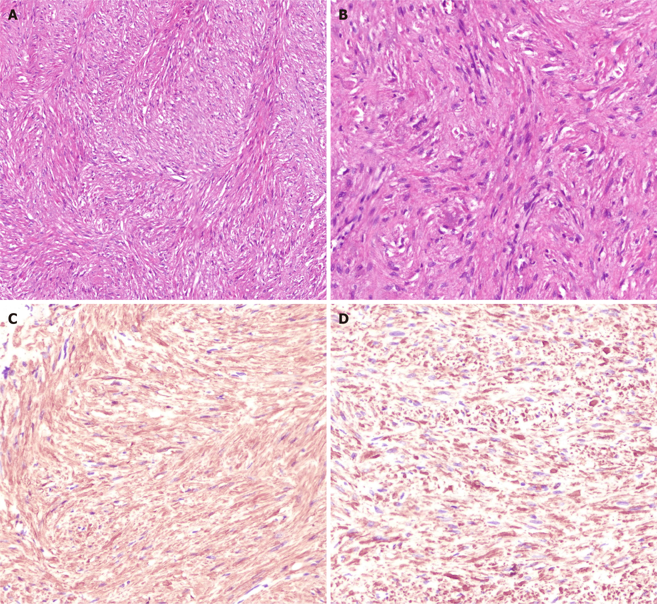
Figure 3 Histopathology and immunohistochemistry suggested leiomyoma.
A: HE staining showed that the tumor cells were long spindle-shaped, and the cytoplasm was rich in red stain and was arranged in bundles or interleaved shapes (HE × 20); B: At high magnification, HE staining showed that the nucleus was long rod-shaped (longitudinal cut) or round oval (transverse cut), located in the center of the cytoplasm, and the nucleolus was not obvious. The nuclear morphology was mild and heterotypic, the chromatin was uniform, and the mitosis was rare (HE × 40); C: Immunohistochemistry showed positive SMA (× 40); D: Immunohistochemistry indicated positive Desmin (× 40).
- Citation: Li P, Tang GM, Li PL, Zhang C, Wang WQ. Endoscopic resection of a giant irregular leiomyoma in fundus and cardia: A case report. World J Gastrointest Endosc 2024; 16(12): 678-685
- URL: https://www.wjgnet.com/1948-5190/full/v16/i12/678.htm
- DOI: https://dx.doi.org/10.4253/wjge.v16.i12.678


