Copyright
©The Author(s) 2024.
World J Gastrointest Endosc. Dec 16, 2024; 16(12): 678-685
Published online Dec 16, 2024. doi: 10.4253/wjge.v16.i12.678
Published online Dec 16, 2024. doi: 10.4253/wjge.v16.i12.678
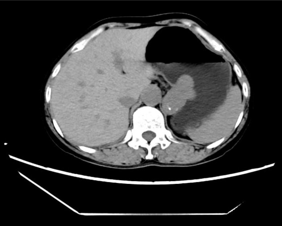
Figure 1 Computed tomography revealed an irregular submucosal mass in the gastric fundus and cardia, some of which protruded out of the cavity, with a size of about 4.
5 cm × 3.2 cm × 4.9 cm and a clear boundary. Nodular calcification was observed within the lesion, with a computed tomography (CT) value of about 42 HU, and mild enhancement was observed. The tertiary CT values were about 52 HU, 54 HU, and 55 HU, and linear enhancement was observed at the lateral edge of the lesion. There were no obvious enlarged lymph nodes around the lesion.
- Citation: Li P, Tang GM, Li PL, Zhang C, Wang WQ. Endoscopic resection of a giant irregular leiomyoma in fundus and cardia: A case report. World J Gastrointest Endosc 2024; 16(12): 678-685
- URL: https://www.wjgnet.com/1948-5190/full/v16/i12/678.htm
- DOI: https://dx.doi.org/10.4253/wjge.v16.i12.678


