Copyright
©The Author(s) 2024.
World J Gastrointest Endosc. Nov 16, 2024; 16(11): 617-622
Published online Nov 16, 2024. doi: 10.4253/wjge.v16.i11.617
Published online Nov 16, 2024. doi: 10.4253/wjge.v16.i11.617
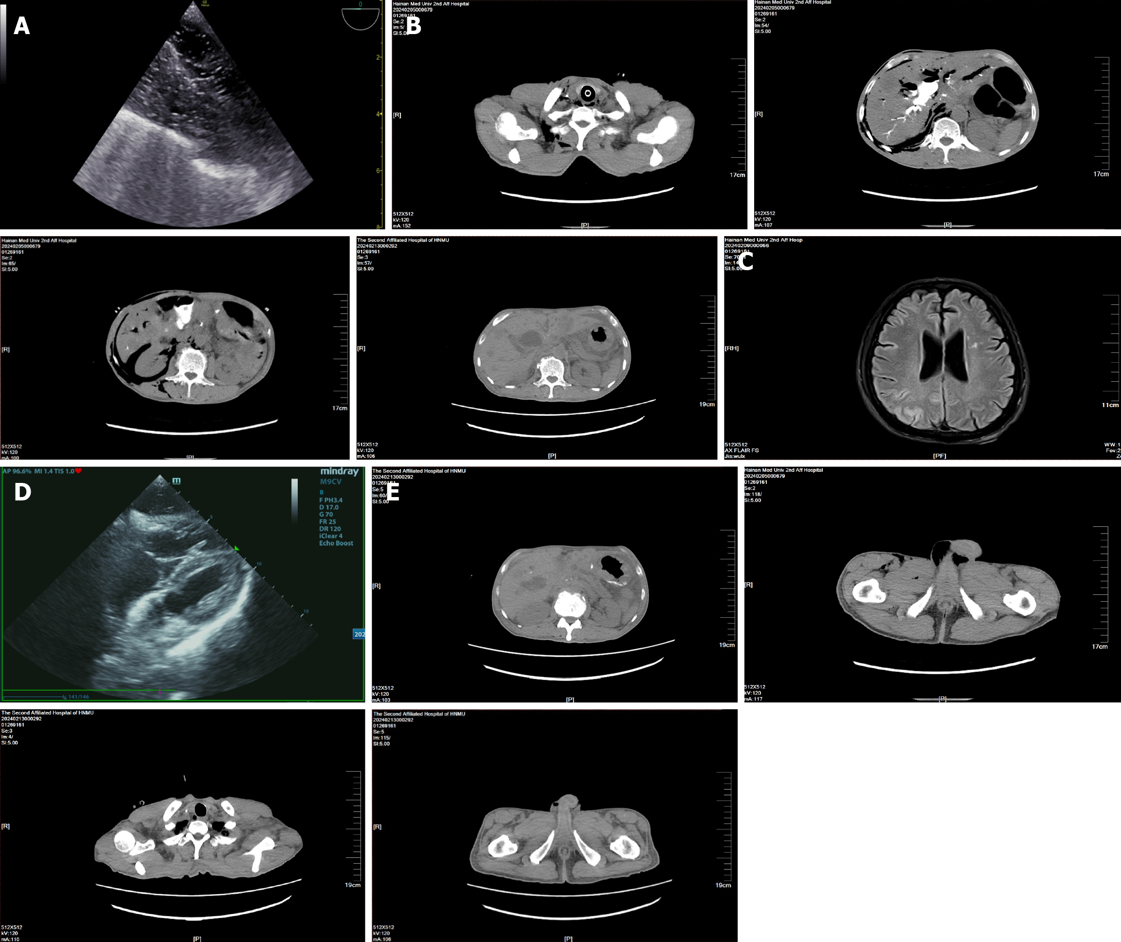
Figure 1 Imaging evolution of systemic pneumatosis in air embolism post-endoscopic retrograde cholangiopancreatography.
A: Esophageal echocardiography suggests intracardiac pneumatosis; B: Pneumatosis is present in the right chest, back and abdominal wall, mediastinum, right pleural cavity, liver parenchyma, adjacent to the inferior vena cava, retroperitoneum, around the right kidney, right groin and scrotum, and proximal right thigh; C: Magnetic resonance imaging of the head indicates ischemic-hypoxic brain injury; D: Echocardiography suggests a reduction in intracardiac pneumatosis compared to previous; E: Pneumatosis in the original areas of the right chest, back and abdominal wall, mediastinum, right pleural cavity, adjacent to the inferior vena cava, retroperitoneum, and around the right kidney has essentially disappeared, while pneumatosis in the right groin and scrotum has decreased compared to previous.
- Citation: Li JH, Luo ZK, Zhang Y, Lu TT, Deng Y, Shu RT, Yu H. Systemic air embolism associated with endoscopic retrograde cholangiopancreatography: A case report. World J Gastrointest Endosc 2024; 16(11): 617-622
- URL: https://www.wjgnet.com/1948-5190/full/v16/i11/617.htm
- DOI: https://dx.doi.org/10.4253/wjge.v16.i11.617


