Copyright
©The Author(s) 2024.
World J Gastrointest Endosc. Oct 16, 2024; 16(10): 545-556
Published online Oct 16, 2024. doi: 10.4253/wjge.v16.i10.545
Published online Oct 16, 2024. doi: 10.4253/wjge.v16.i10.545
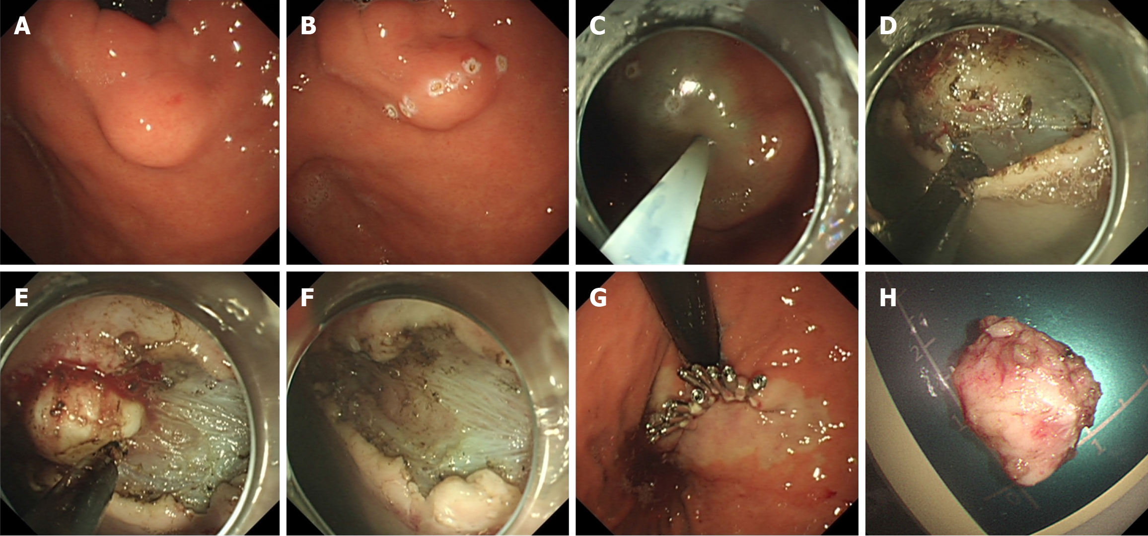
Figure 1 Endoscopic submucosal excavation for treatment of small gastric mesenchymal tumors.
A: Small gastric mesenchymal tumor (sGMT) located at the gastric fundus near the cardia; B: Electrocautery markings visible on the sGMT’s surface; C: Submucosal injection around the sGMT; D: Incision of the mucosal surface using a mucosal incision knife (MIK) to expose the tumor; E: Separation of the tumor from its base using a MIK; F: The wound after complete tumor dissection; G: Closure of the wound using titanium clips; H: The completely resected tumor.
- Citation: Lin XM, Peng YM, Zeng HT, Yang JX, Xu ZL. Endoscopic “calabash” ligation and resection for small gastric mesenchymal tumors. World J Gastrointest Endosc 2024; 16(10): 545-556
- URL: https://www.wjgnet.com/1948-5190/full/v16/i10/545.htm
- DOI: https://dx.doi.org/10.4253/wjge.v16.i10.545


