Copyright
©The Author(s) 2022.
World J Gastrointest Endosc. Sep 16, 2022; 14(9): 536-546
Published online Sep 16, 2022. doi: 10.4253/wjge.v14.i9.536
Published online Sep 16, 2022. doi: 10.4253/wjge.v14.i9.536
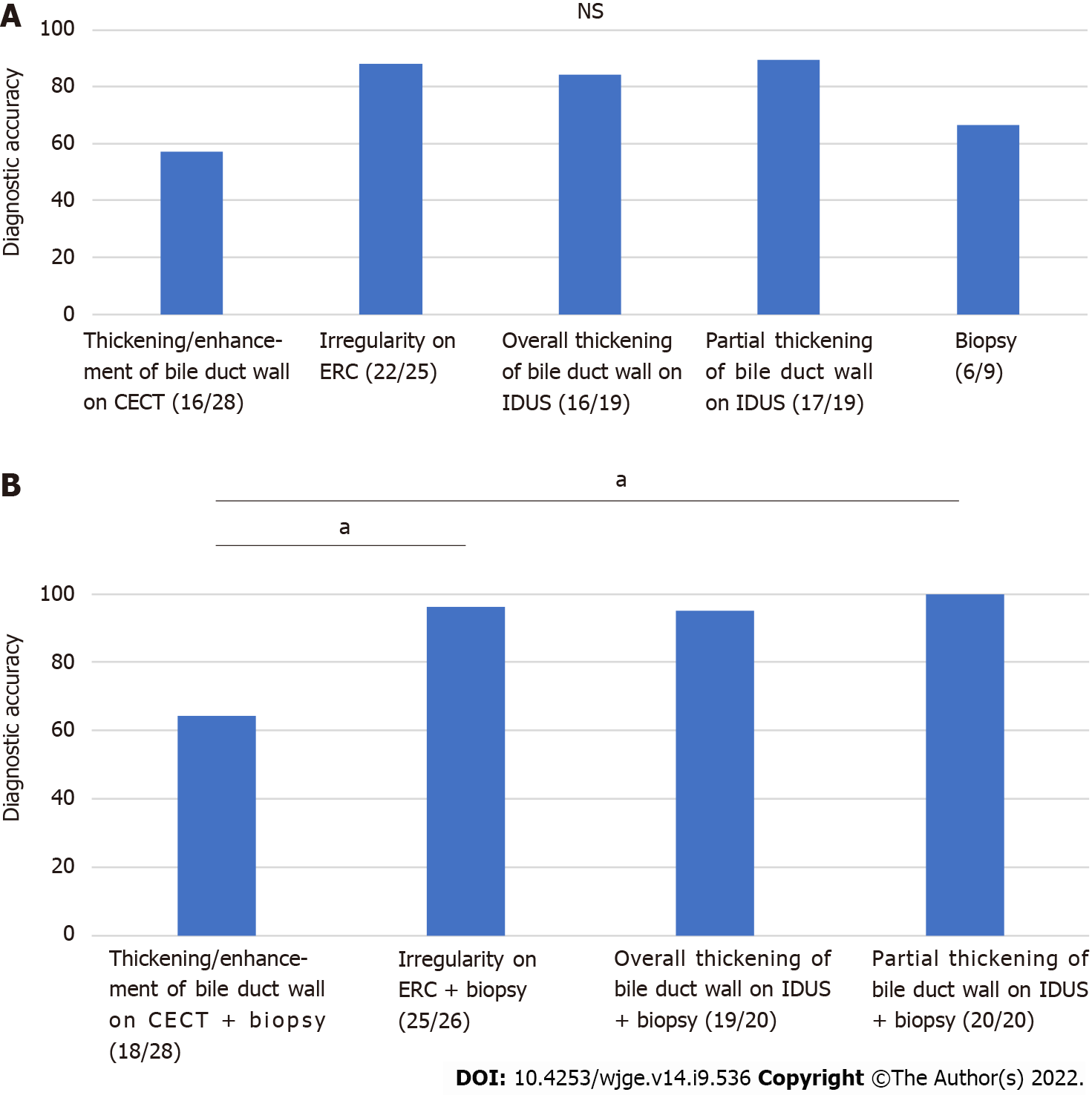
Figure 3 Comparison of methods for diagnosing hilar biliary invasion of ampullary cancer in patients without biliary stents.
A: Partial thickening of the bile duct wall on IDUS showed the highest diagnostic accuracy; B: Among the various combinations (imaging findings and biliary biopsy results) for diagnosing hilar biliary invasion, partial thickening of the bile duct wall on IDUS + biliary biopsy results showed the highest diagnostic accuracy. aP < 0.05. CECT: Contrast-enhanced computed tomography; ERC: Endoscopic retrograde cholangiography; IDUS: Intraductal ultrasonography; NS: Not significant.
- Citation: Takagi T, Sugimoto M, Suzuki R, Konno N, Asama H, Sato Y, Irie H, Nakamura J, Takasumi M, Hashimoto M, Kato T, Kobashi R, Yanagita T, Hashimoto Y, Marubashi S, Hikichi T, Ohira H. Screening for hilar biliary invasion in ampullary cancer patients. World J Gastrointest Endosc 2022; 14(9): 536-546
- URL: https://www.wjgnet.com/1948-5190/full/v14/i9/536.htm
- DOI: https://dx.doi.org/10.4253/wjge.v14.i9.536


