Copyright
©The Author(s) 2022.
World J Gastrointest Endosc. Jul 16, 2022; 14(7): 455-466
Published online Jul 16, 2022. doi: 10.4253/wjge.v14.i7.455
Published online Jul 16, 2022. doi: 10.4253/wjge.v14.i7.455
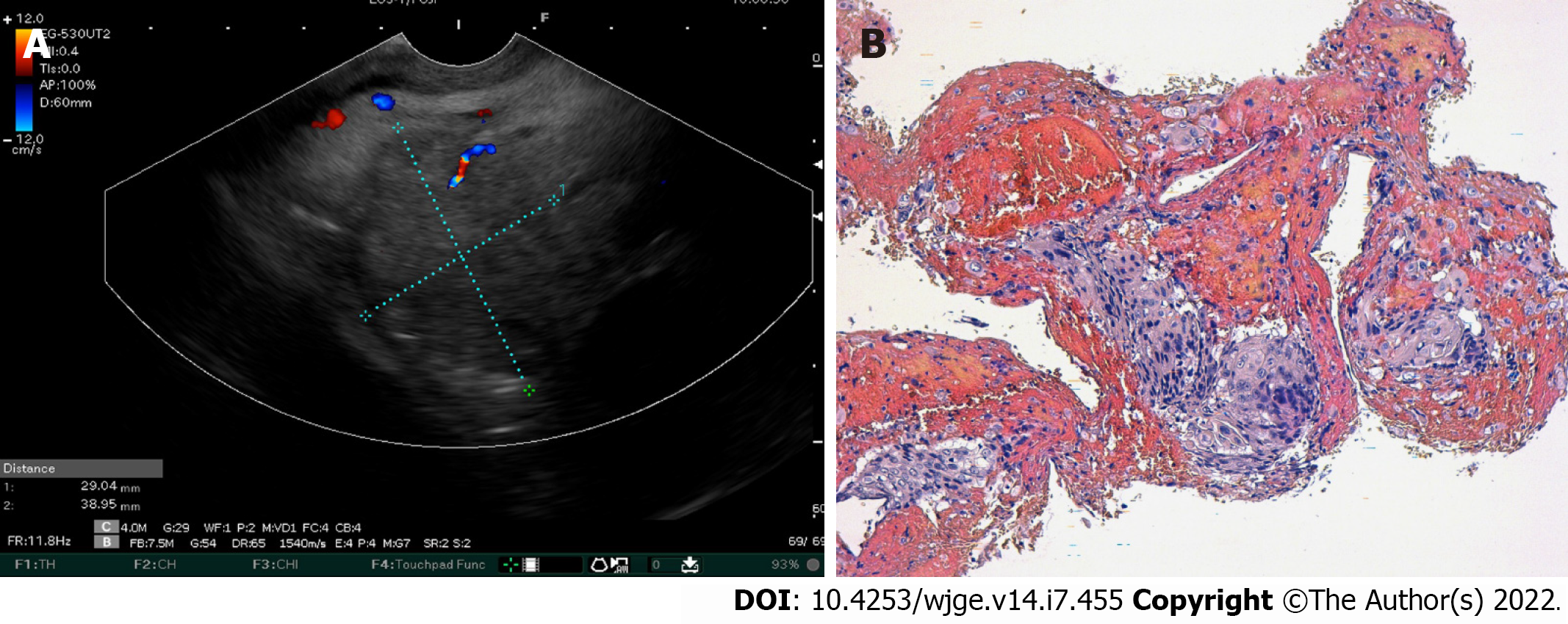
Figure 3 Images of endoscopic ultrasound and histological analysis of the pancreatic mass.
A: Linear endoscopic ultrasound showed a pancreatic head tumor; B: Microphotography showing a proliferation with an easily recognizable squamous differentiation, including apparent intercellular bridges and minimal pleomorphism. Hematoxylin-eosin stain (× 200).
- Citation: Rais K, El Eulj O, El Moutaoukil N, Kamaoui I, Bennani A, Kharrasse G, Zazour A, Khannoussi W, Ismaili Z. Solitary pancreatic metastasis from squamous cell lung carcinoma: A case report and review of literature. World J Gastrointest Endosc 2022; 14(7): 455-466
- URL: https://www.wjgnet.com/1948-5190/full/v14/i7/455.htm
- DOI: https://dx.doi.org/10.4253/wjge.v14.i7.455


