Copyright
©The Author(s) 2022.
World J Gastrointest Endosc. May 16, 2022; 14(5): 320-334
Published online May 16, 2022. doi: 10.4253/wjge.v14.i5.320
Published online May 16, 2022. doi: 10.4253/wjge.v14.i5.320
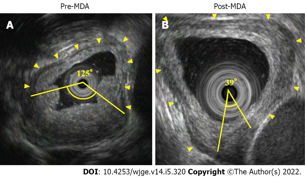
Figure 4 Measurements of muscle layer defect angle.
A: Endoscopic ultrasound showed the normal muscle layer as hypoechoic inner muscle layer, hyperechoic intermuscular connective tissue layer, and hypoechoic outer muscle layer (arrowhead). In this case of cT3 before neoadjuvant chemotherapy (NAC), pre-muscle layer defect angle (MDA) was 125°; B: After NAC, post-MDA was 39°, and thus MDA reduction rate was 34.8%. This case achieved pCR.
- Citation: Yonemoto S, Uesato M, Nakano A, Murakami K, Toyozumi T, Maruyama T, Suito H, Tamachi T, Kato M, Kainuma S, Matsusaka K, Matsubara H. Why is endosonography insufficient for residual diagnosis after neoadjuvant therapy for esophageal cancer? Solutions using muscle layer evaluation. World J Gastrointest Endosc 2022; 14(5): 320-334
- URL: https://www.wjgnet.com/1948-5190/full/v14/i5/320.htm
- DOI: https://dx.doi.org/10.4253/wjge.v14.i5.320


