Copyright
©The Author(s) 2022.
World J Gastrointest Endosc. Apr 16, 2022; 14(4): 191-204
Published online Apr 16, 2022. doi: 10.4253/wjge.v14.i4.191
Published online Apr 16, 2022. doi: 10.4253/wjge.v14.i4.191
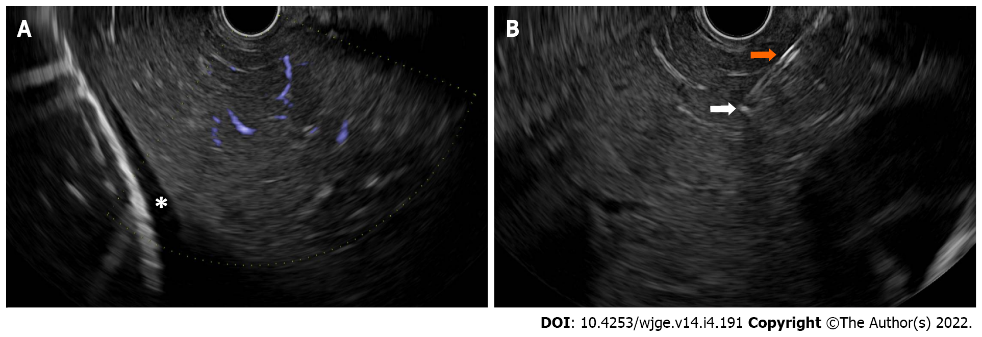
Figure 4 Endoscopic ultrasound guided liver biopsy.
A: Liver parenchyma without major intervening intrahepatic blood vessels, which is an optimal location for endoscopic ultrasound-guided liver biopsy. An asterisk indicates a small amount of perihepatic ascites; B: An endoscopic ultrasound-guided liver biopsy using a heparin-primed wet-suction technique via a 19-gauge Franseen needle tip design. The hyperechoic tip of the needle (white arrow) and the shaft of the needle (orange arrow) must be visualized at all times during the fine needle biopsy of the liver.
- Citation: Kerdsirichairat T, Shin EJ. Endoscopic ultrasound guided interventions in the management of pancreatic cancer. World J Gastrointest Endosc 2022; 14(4): 191-204
- URL: https://www.wjgnet.com/1948-5190/full/v14/i4/191.htm
- DOI: https://dx.doi.org/10.4253/wjge.v14.i4.191


