Copyright
©The Author(s) 2022.
World J Gastrointest Endosc. Mar 16, 2022; 14(3): 129-141
Published online Mar 16, 2022. doi: 10.4253/wjge.v14.i3.129
Published online Mar 16, 2022. doi: 10.4253/wjge.v14.i3.129
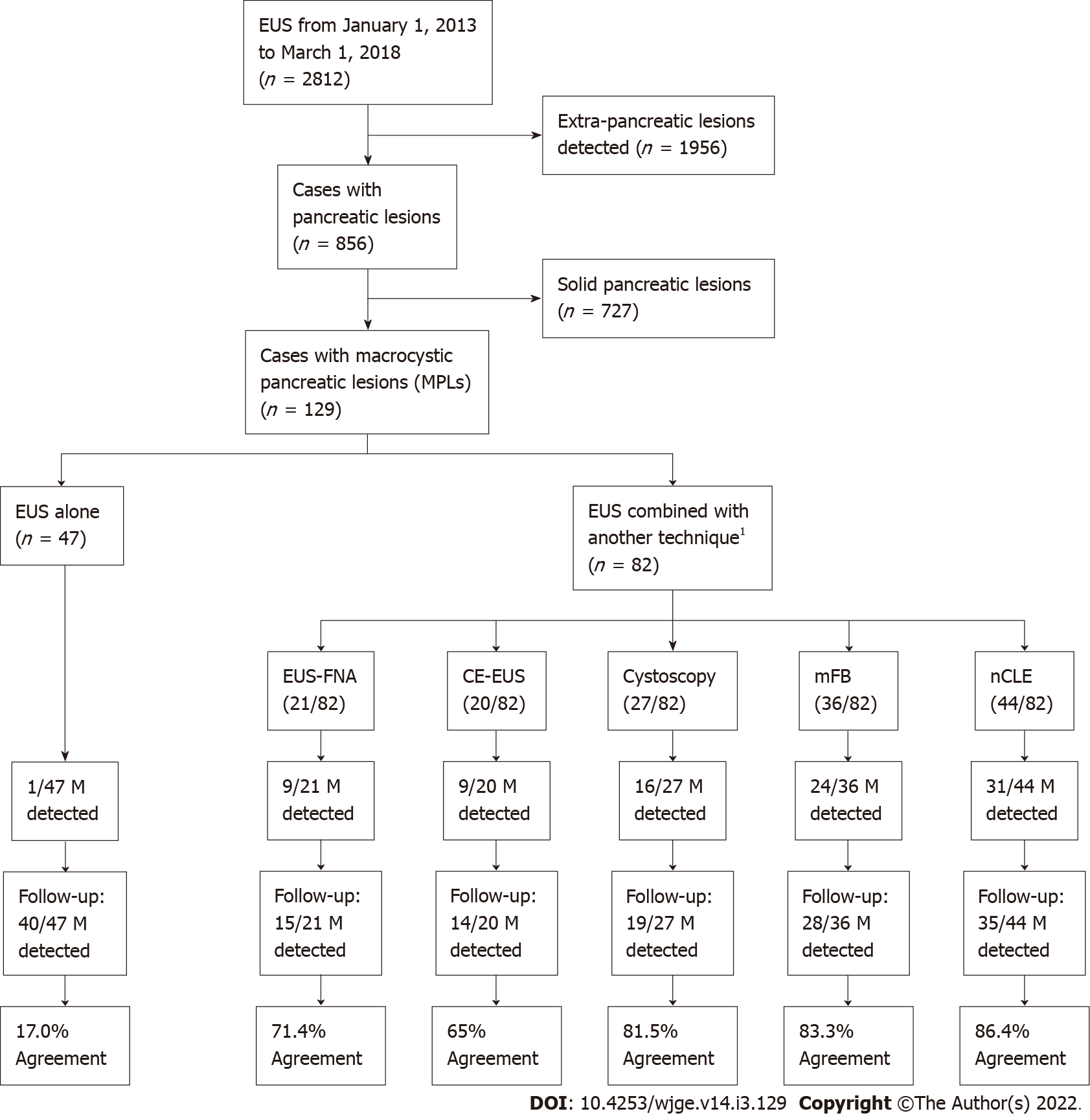
Figure 2 Population study flowchart.
1Numbers of techniques were not mutually exclusive. Endoscopic ultrasound could be combined with more than one other technique, as shown on the illustrated Venn diagram in Figure 3. EUS: Endoscopic ultrasound; EUS-FNA: Endoscopic ultrasound-guided fine needle aspiration; Cystoscopy: Fiberoptic probe cystoscopy; nCLE: Endoscopic ultrasound-guided needle-based confocal laser-endomicroscopy; mFB: Endoscopic ultrasound-guided through-the-needle direct intracystic micro forceps biopsy; CE-EUS: Contrast-enhanced endoscopic ultrasound; M: Malignancy.
- Citation: Robles-Medranda C, Olmos JI, Puga-Tejada M, Oleas R, Baquerizo-Burgos J, Arevalo-Mora M, Del Valle Zavala R, Nebel JA, Calle Loffredo D, Pitanga-Lukashok H. Endoscopic ultrasound-guided through-the-needle microforceps biopsy and needle-based confocal laser-endomicroscopy increase detection of potentially malignant pancreatic cystic lesions: A single-center study. World J Gastrointest Endosc 2022; 14(3): 129-141
- URL: https://www.wjgnet.com/1948-5190/full/v14/i3/129.htm
- DOI: https://dx.doi.org/10.4253/wjge.v14.i3.129


