Copyright
©The Author(s) 2022.
World J Gastrointest Endosc. Dec 16, 2022; 14(12): 789-794
Published online Dec 16, 2022. doi: 10.4253/wjge.v14.i12.789
Published online Dec 16, 2022. doi: 10.4253/wjge.v14.i12.789
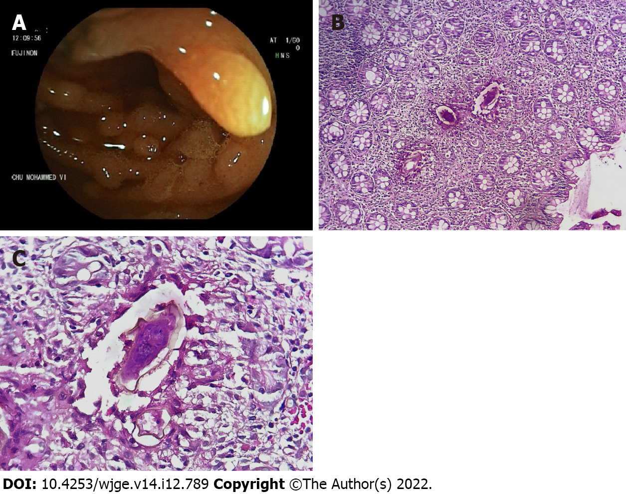
Figure 1 Colonoscopy and histopathological findings.
A: Polyps were observed during colonoscopy; B: Microphotography showed the presence of three Schistosoma eggs in the colic mucosa (hematoxylin and eosin, × 40); C: Microphotography of a Schistosoma egg showed a thick peripheral capsule and a viable embryo inside. The egg capsule was surrounded by numerous eosinophils (hematoxylin and eosin, × 400).
- Citation: Koulali H, Zazour A, Khannoussi W, Kharrasse G, Ismaili Z. Colonic schistosomiasis: A case report. World J Gastrointest Endosc 2022; 14(12): 789-794
- URL: https://www.wjgnet.com/1948-5190/full/v14/i12/789.htm
- DOI: https://dx.doi.org/10.4253/wjge.v14.i12.789


