Copyright
©The Author(s) 2022.
World J Gastrointest Endosc. Oct 16, 2022; 14(10): 642-647
Published online Oct 16, 2022. doi: 10.4253/wjge.v14.i10.642
Published online Oct 16, 2022. doi: 10.4253/wjge.v14.i10.642
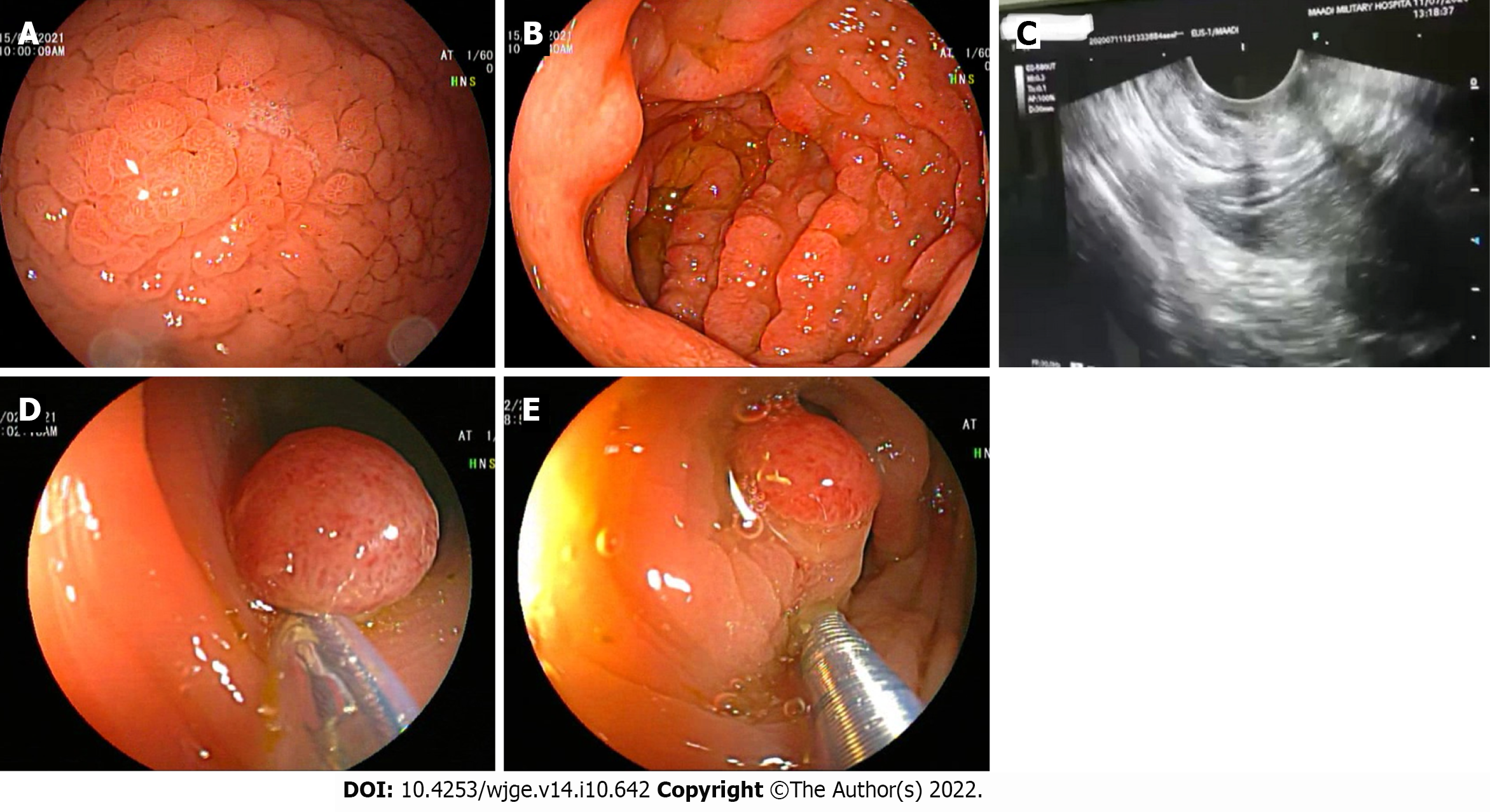
Figure 2 Endoscopy.
A and B: Upper endoscopy revealed a diffuse markedly thickened gastric mucosa with numerous polypoidal lesions; C: Endoscopic ultrasound revealed a significantly hypertrophic mucosa and muscularis mucosa, but sparing of the submucosa and the muscularis propria; D and E: Colonoscopy showed multiple variable-sized, sessile, and pedunculated polyps, which were removed by snare polypectomy.
- Citation: Alzamzamy AE, Aboubakr A, Okasha HH, Abdellatef A, Elkholy S, Wahba M, Alboraie M, Elsayed H, Othman MO. Cronkhite-Canada syndrome: First case report from Egypt and North Africa. World J Gastrointest Endosc 2022; 14(10): 642-647
- URL: https://www.wjgnet.com/1948-5190/full/v14/i10/642.htm
- DOI: https://dx.doi.org/10.4253/wjge.v14.i10.642


