Copyright
©The Author(s) 2021.
World J Gastrointest Endosc. Sep 16, 2021; 13(9): 437-446
Published online Sep 16, 2021. doi: 10.4253/wjge.v13.i9.437
Published online Sep 16, 2021. doi: 10.4253/wjge.v13.i9.437
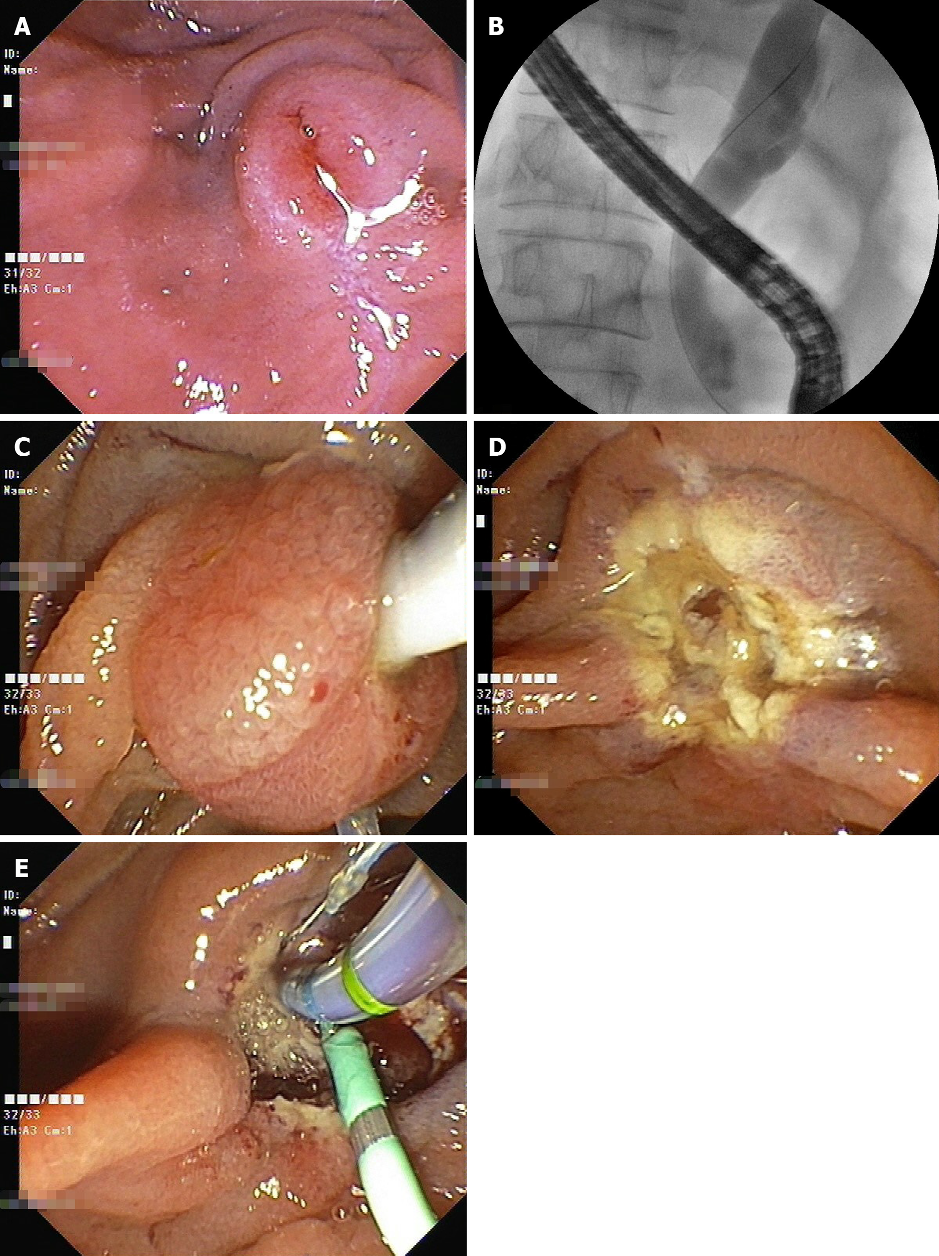
Figure 2 Endoscopic snare papillectomy.
A: Endoscopic view of the sub-epithelial ampullary lesion; B: Cholangiogram showing terminal common bile duct (CBD) stricture with upstream dilated CBD after selective CBD cannulation; C: Endoscopic view showing snaring of the papilla while pulling back the expanded balloon within the CBD towards the duodenal lumen; D: Endoscopic view after endoscopic snare papillectomy; E: Endoscopic view of the biliary sphincterotomy and pancreatic stent in place.
- Citation: Vyawahare MA, Musthyla NB. Ectopic pancreas at the ampulla of Vater diagnosed with endoscopic snare papillectomy: A case report and review of literature . World J Gastrointest Endosc 2021; 13(9): 437-446
- URL: https://www.wjgnet.com/1948-5190/full/v13/i9/437.htm
- DOI: https://dx.doi.org/10.4253/wjge.v13.i9.437


