Copyright
©The Author(s) 2021.
World J Gastrointest Endosc. Jul 16, 2021; 13(7): 233-237
Published online Jul 16, 2021. doi: 10.4253/wjge.v13.i7.233
Published online Jul 16, 2021. doi: 10.4253/wjge.v13.i7.233
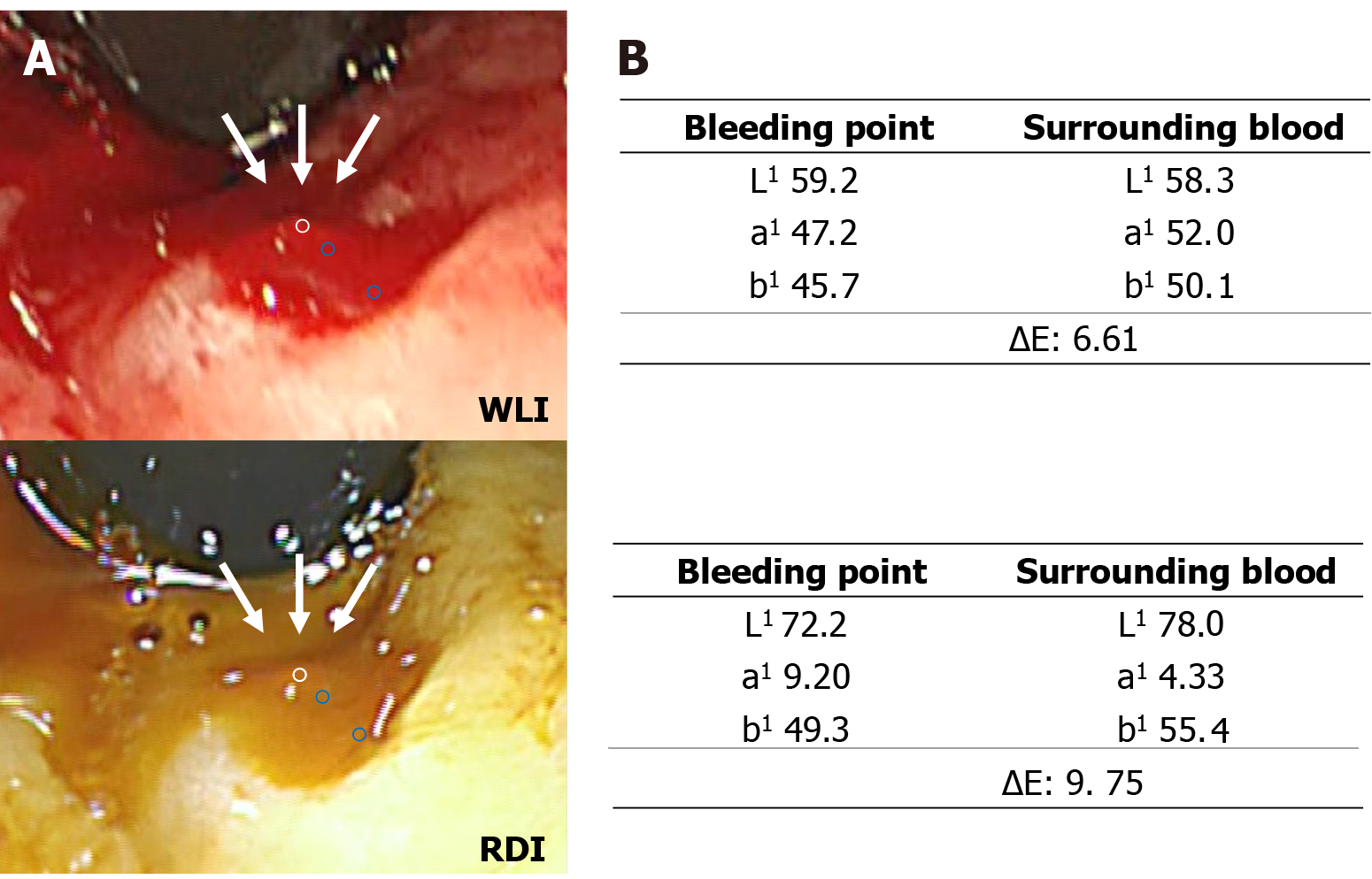
Figure 3 Color values and color differences between bleeding point and surrounding blood.
A: The regions of interests (ROIs) were located in the bleeding point and at two selected points in surrounding blood (one was just next to outside of the bleeding point and the other was just inside the surrounding blood), avoiding areas with halation. Each ROI was set approximately in the same region for white light imaging and red dichromatic imaging. The white and blue circles indicate the ROI of the bleeding point (white arrow) and surrounding blood, respectively. The color values were defined as the median color value in each ROI; and B: The ΔE based on color value change between the ROI of the bleeding point and surrounding blood. WLI: White light imaging; RDI: Red dichromatic imaging; ΔE: Color difference.
- Citation: Hirai Y, Kayashima A, Nakazato Y, Fujimoto A. Visibility of the bleeding point in acute rectal hemorrhagic ulcer using red dichromatic imaging: A case report. World J Gastrointest Endosc 2021; 13(7): 233-237
- URL: https://www.wjgnet.com/1948-5190/full/v13/i7/233.htm
- DOI: https://dx.doi.org/10.4253/wjge.v13.i7.233


