Copyright
©The Author(s) 2021.
World J Gastrointest Endosc. Dec 16, 2021; 13(12): 649-658
Published online Dec 16, 2021. doi: 10.4253/wjge.v13.i12.649
Published online Dec 16, 2021. doi: 10.4253/wjge.v13.i12.649
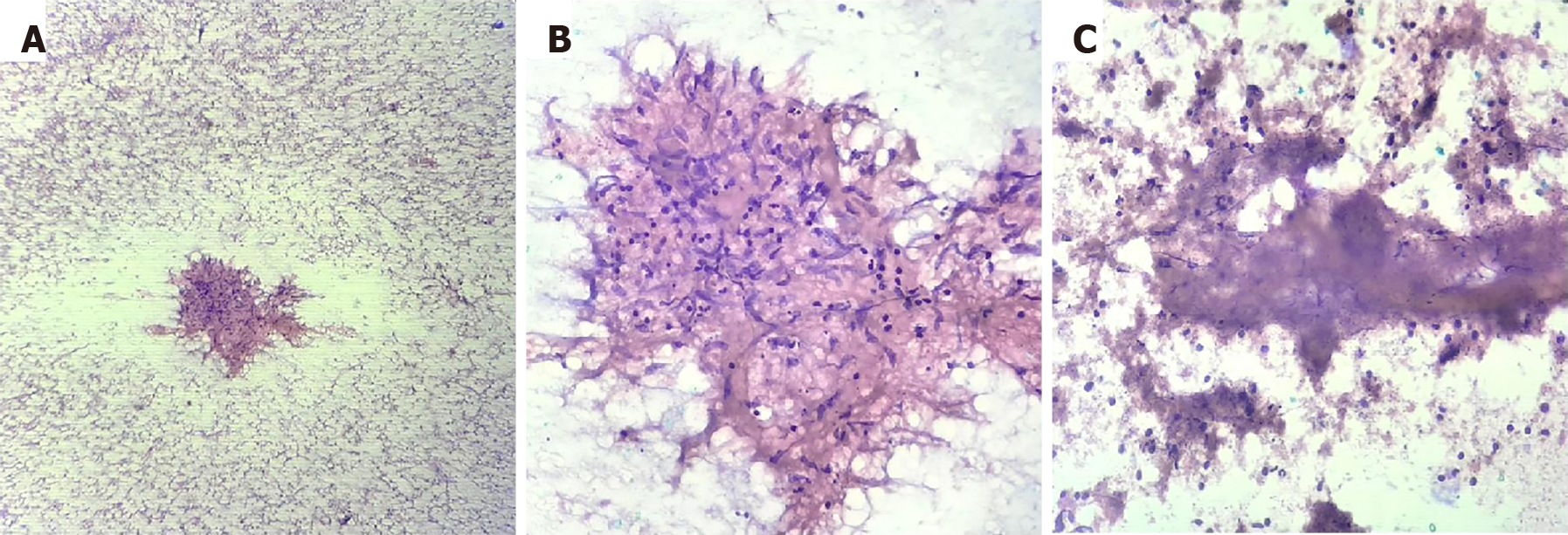
Figure 1 Histopathology findings on the fine needle aspiration sample of a patient with tuberculosis.
A: An epithelioid cell granuloma with scattered lymphocytes and red blood cells in the background (100 ×); B: Collection of epithelioid histiocytes forming a granuloma (400 ×); C: Necrotic material and inflammatory cells (400 ×).
- Citation: Rao B H, Nair P, Priya SK, Vallonthaiel AG, Sathyapalan DT, Koshy AK, Venu RP. Role of endoscopic ultrasound guided fine needle aspiration/biopsy in the evaluation of intra-abdominal lymphadenopathy due to tuberculosis. World J Gastrointest Endosc 2021; 13(12): 649-658
- URL: https://www.wjgnet.com/1948-5190/full/v13/i12/649.htm
- DOI: https://dx.doi.org/10.4253/wjge.v13.i12.649


