Copyright
©The Author(s) 2021.
World J Gastrointest Endosc. Oct 16, 2021; 13(10): 529-542
Published online Oct 16, 2021. doi: 10.4253/wjge.v13.i10.529
Published online Oct 16, 2021. doi: 10.4253/wjge.v13.i10.529
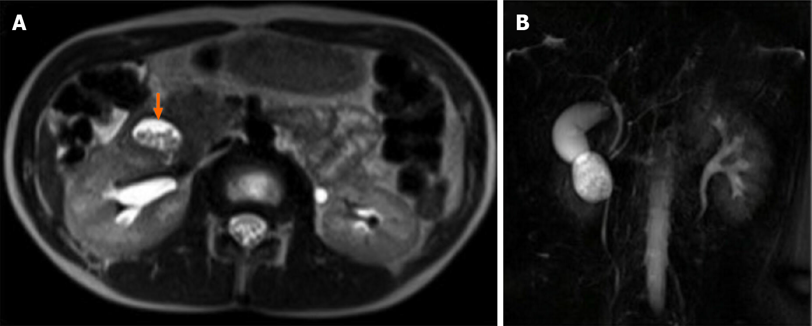
Figure 5 Magnetic resonance imaging.
A: Oval mass is located below the gallbladder and lateral to the common bile duct and pancreatic duct, adjacent to the pancreatic head. The cyst was filled with fluid and multiple stones; B: Cholangiographic reconstruction showed normal gallbladder and intra- and extrahepatic bile ducts.
- Citation: Bulotta AL, Stern MV, Moneghini D, Parolini F, Bondioni MP, Missale G, Boroni G, Alberti D. Endoscopic treatment of periampullary duodenal duplication cysts in children: Four case reports and review of the literature. World J Gastrointest Endosc 2021; 13(10): 529-542
- URL: https://www.wjgnet.com/1948-5190/full/v13/i10/529.htm
- DOI: https://dx.doi.org/10.4253/wjge.v13.i10.529


