Copyright
©The Author(s) 2021.
World J Gastrointest Endosc. Oct 16, 2021; 13(10): 529-542
Published online Oct 16, 2021. doi: 10.4253/wjge.v13.i10.529
Published online Oct 16, 2021. doi: 10.4253/wjge.v13.i10.529
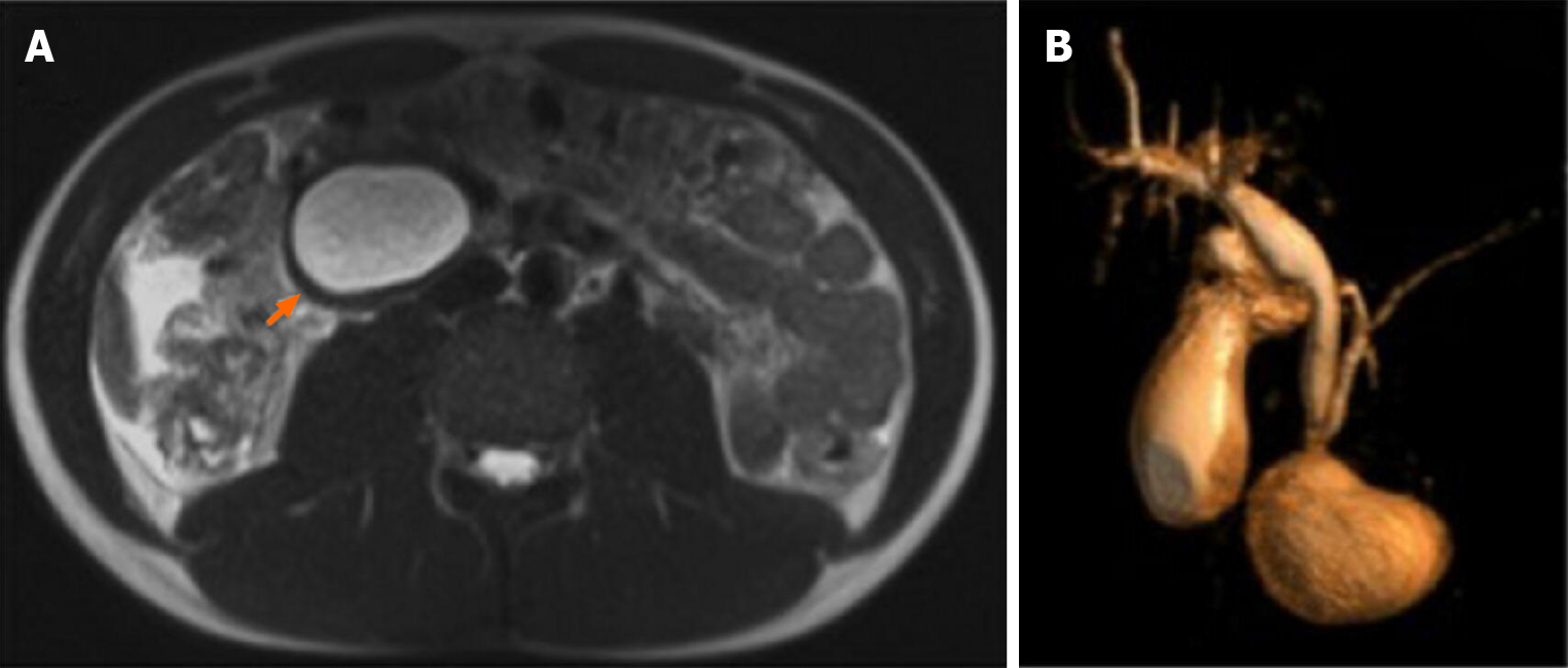
Figure 1 Magnetic resonance imaging on HASTE T2 w sequence.
A: Homogeneously hyperintense cyst located within the duodenum, which was partially occluded (arrow); B: On 3D cholangiographic reconstruction, intrahepatic bile ducts were normal, cystic duct was dilated with tortuous course and common hepatic duct presented saccular dilation. Common bile duct had a caliber at the upper limits of the normal range with a regular course and was in communication with periampullary duodenal duplication cysts.
- Citation: Bulotta AL, Stern MV, Moneghini D, Parolini F, Bondioni MP, Missale G, Boroni G, Alberti D. Endoscopic treatment of periampullary duodenal duplication cysts in children: Four case reports and review of the literature. World J Gastrointest Endosc 2021; 13(10): 529-542
- URL: https://www.wjgnet.com/1948-5190/full/v13/i10/529.htm
- DOI: https://dx.doi.org/10.4253/wjge.v13.i10.529


