Copyright
©The Author(s) 2021.
World J Gastrointest Endosc. Oct 16, 2021; 13(10): 518-528
Published online Oct 16, 2021. doi: 10.4253/wjge.v13.i10.518
Published online Oct 16, 2021. doi: 10.4253/wjge.v13.i10.518
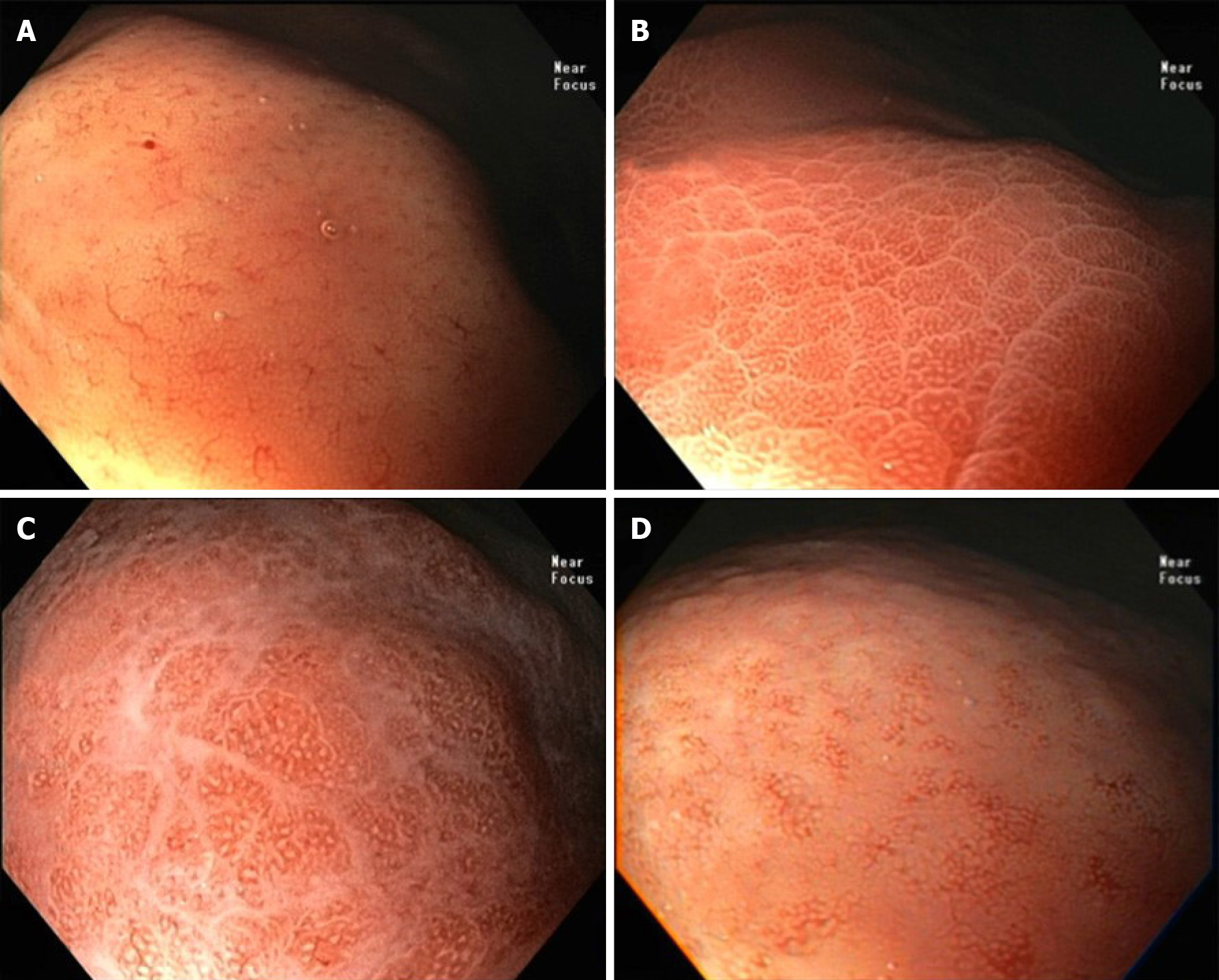
Figure 2 Near focus examination of gastric body.
A: Type 1: regular arrangement of collecting venules and regular round pits; B: Type 2: regular round pits, with erythema, sulci and loss of collecting venules; C: Type 3: loss of normal subepithelial capillary network (SECN) and collecting venules and with white enlarged pits surrounded by erythema and exudate; D: Type 4: loss of normal SECN and round pits, with irregular arrangement of collecting venules.
- Citation: Fiuza F, Maluf-Filho F, Ide E, Furuya Jr CK, Fylyk SN, Ruas JN, Stabach L, Araujo GA, Matuguma SE, Uemura RS, Sakai CM, Yamazaki K, Ueda SS, Sakai P, Martins BC. Association between mucosal surface pattern under near focus technology and Helicobacter pylori infection. World J Gastrointest Endosc 2021; 13(10): 518-528
- URL: https://www.wjgnet.com/1948-5190/full/v13/i10/518.htm
- DOI: https://dx.doi.org/10.4253/wjge.v13.i10.518


