Copyright
©The Author(s) 2020.
World J Gastrointest Endosc. Sep 16, 2020; 12(9): 304-309
Published online Sep 16, 2020. doi: 10.4253/wjge.v12.i9.304
Published online Sep 16, 2020. doi: 10.4253/wjge.v12.i9.304
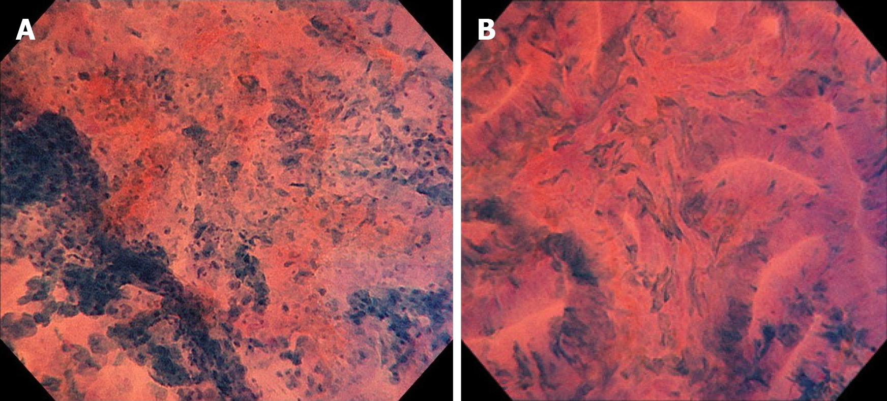
Figure 2 Endocytoscopy of the lesion.
A: On the left side of the lesion (depression), unclear gland formation and agglomeration of distorted nuclei strongly stained by methylene blue were visible with endocytoscopy, suggesting a submucosal invasive cancer; B: On the right side of the lesion (flat area), slit-like smooth lumens and a regular pattern of fusiform nuclei were found, suggesting an adenoma.
- Citation: Akimoto Y, Kudo SE, Ichimasa K, Kouyama Y, Misawa M, Hisayuki T, Kudo T, Nemoto T. Small invasive colon cancer with adenoma observed by endocytoscopy: A case report. World J Gastrointest Endosc 2020; 12(9): 304-309
- URL: https://www.wjgnet.com/1948-5190/full/v12/i9/304.htm
- DOI: https://dx.doi.org/10.4253/wjge.v12.i9.304


