Copyright
©The Author(s) 2020.
World J Gastrointest Endosc. Oct 16, 2020; 12(10): 355-364
Published online Oct 16, 2020. doi: 10.4253/wjge.v12.i10.355
Published online Oct 16, 2020. doi: 10.4253/wjge.v12.i10.355
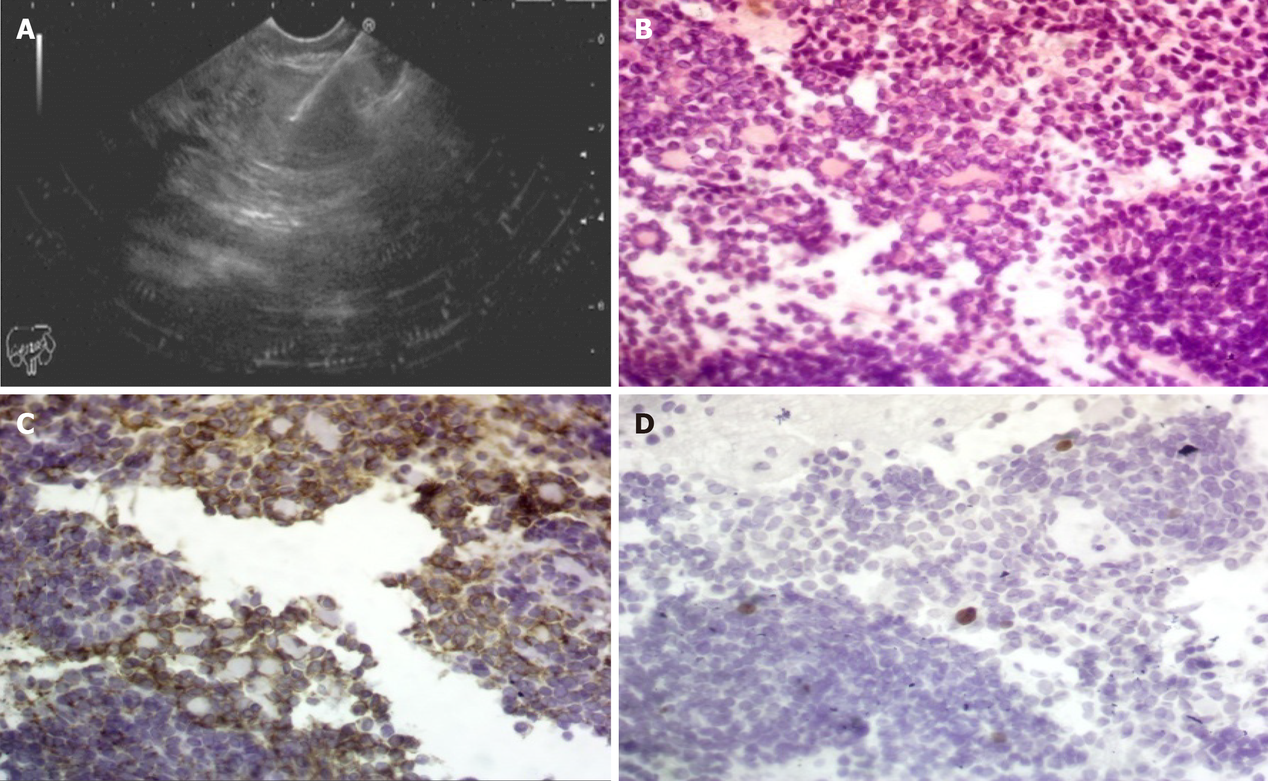
Figure 1 Endoscopic ultrasound and hematoxylin/eosin staining.
A: Pancreatic head mass with fine needle aspiration; B: Hematoxylin/eosin staining: Shows cellular tumor tissue formed by small cells with focal resetting and tumor cell nuclei show fine chromatin with a little cytoplasm (Hematoxylin/eosin, 400 ×); C: Shows moderate membranous reaction of the tumor cells (CD56, 400 ×); and D: Show positive nuclear staining in a few tumor cells (< 2%) (Ki-67, 400 ×); consistent with a well-differentiated neuroendocrine tumor.
- Citation: Altonbary AY, Hakim H, Elkashef W. Role of endoscopic ultrasound in pediatric patients: A single tertiary center experience and review of the literature. World J Gastrointest Endosc 2020; 12(10): 355-364
- URL: https://www.wjgnet.com/1948-5190/full/v12/i10/355.htm
- DOI: https://dx.doi.org/10.4253/wjge.v12.i10.355


