Copyright
©The Author(s) 2019.
World J Gastrointest Endosc. May 16, 2019; 11(5): 383-388
Published online May 16, 2019. doi: 10.4253/wjge.v11.i5.383
Published online May 16, 2019. doi: 10.4253/wjge.v11.i5.383
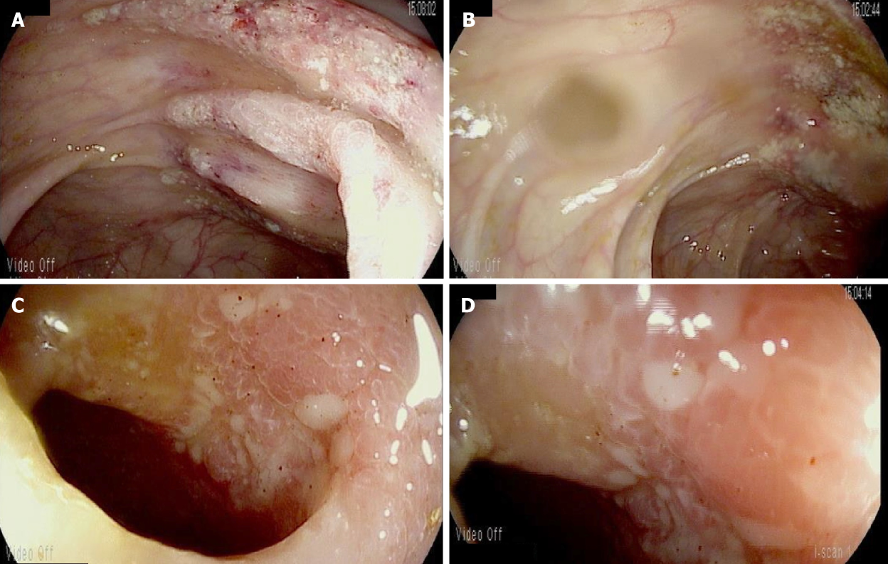
Figure 2 Colonoscopy and ileoscopy findings for case 1.
A, B: For our first case, colonoscopy shows granular erythematous mucosa of the ascending colon (A and B) distal to the ileocolonic anastomosis; C, D: On ileoscopy, evidence of ulceration can be seen in the distal ileum (C and D).
- Citation: Dao AE, Hsu A, Nakshabandi A, Mandaliya R, Nadella S, Sivaraman A, Mattar M, Charabaty A. Role of colonoscopy in diagnosis of capecitabine associated ileitis: Two case reports. World J Gastrointest Endosc 2019; 11(5): 383-388
- URL: https://www.wjgnet.com/1948-5190/full/v11/i5/383.htm
- DOI: https://dx.doi.org/10.4253/wjge.v11.i5.383


