Copyright
©The Author(s) 2019.
World J Gastrointest Endosc. May 16, 2019; 11(5): 373-382
Published online May 16, 2019. doi: 10.4253/wjge.v11.i5.373
Published online May 16, 2019. doi: 10.4253/wjge.v11.i5.373
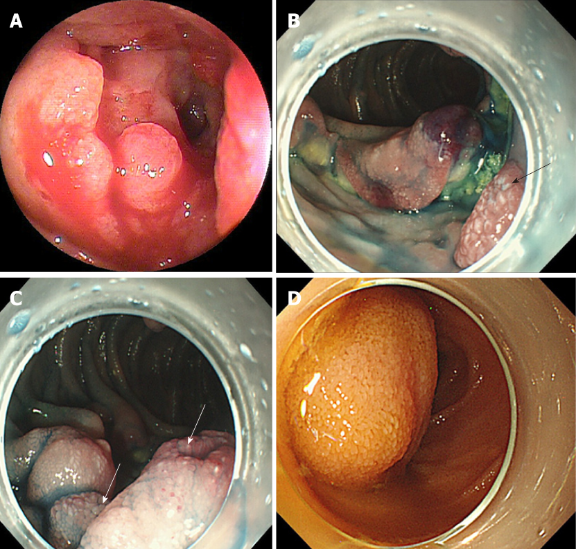
Figure 2 Endoscopic findings for each small intestinal tumor.
A: Representative image of an epithelial tumor (Group 1: primary small intestinal cancer). This tumor was solitary and located in the jejunum. The type was infiltrative ulcerated type. This tumor was also associated with stenosis and bleeding; B and C: Representative images of malignant lymphoma (Group 2: diffuse large B-cell lymphoma). These tumors were multiple and located in the jejunum and ileum and appeared as ulcerated masses with raised margins. These tumors also had white villi (arrows) and were not associated with stenosis or bleeding; D: Representative image of a gastrointestinal stromal tumor (Group 3). This tumor was solitary and located in the jejunum and appeared as a protruded mass. This tumor was not associated with stenosis or bleeding.
- Citation: Horie T, Hosoe N, Takabayashi K, Hayashi Y, Kamiya KJL, Miyanaga R, Mizuno S, Fukuhara K, Fukuhara S, Naganuma M, Shimoda M, Ogata H, Kanai T. Endoscopic characteristics of small intestinal malignant tumors observed by balloon-assisted enteroscopy. World J Gastrointest Endosc 2019; 11(5): 373-382
- URL: https://www.wjgnet.com/1948-5190/full/v11/i5/373.htm
- DOI: https://dx.doi.org/10.4253/wjge.v11.i5.373


