Copyright
©The Author(s) 2018.
World J Gastrointest Endosc. Sep 16, 2018; 10(9): 175-183
Published online Sep 16, 2018. doi: 10.4253/wjge.v10.i9.175
Published online Sep 16, 2018. doi: 10.4253/wjge.v10.i9.175
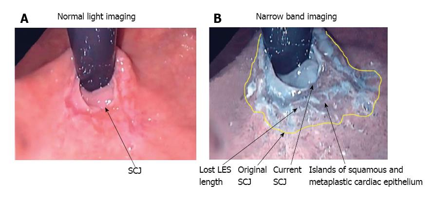
Figure 1 Retroflex endoscopic view of the squamocolumnar junction in advanced gastroesophageal reflux disease.
A: Normal white light image displays a slightly irregular SCJ with normal squamous epithelium extending up the esophagus; B: Narrow band image shows multiple islands of squamous epithelium below the SCJ, surrounded by newly formed metaplastic cardiac epithelium. The splayed out original SCJ (indicated by the yellow line), and damaged portion of the LES between the original SCJ and the current SCJ, take on the appearance of stomach. Loss of esophageal muscle due to inflammation results in a reduction in the abdominal length of the LES and loss of the LES barrier function. Images used with permission from Dr. Peters, Case Western University, Cleveland, OH, United States. GERD: Gastroesophageal reflux disease; LES: Lower esophageal sphincter; SCJ: Squamocolumnar junction.
- Citation: Labenz J, Chandrasoma PT, Knapp LJ, DeMeester TR. Proposed approach to the challenging management of progressive gastroesophageal reflux disease. World J Gastrointest Endosc 2018; 10(9): 175-183
- URL: https://www.wjgnet.com/1948-5190/full/v10/i9/175.htm
- DOI: https://dx.doi.org/10.4253/wjge.v10.i9.175


