Copyright
©The Author(s) 2018.
World J Gastrointest Endosc. Jun 16, 2018; 10(6): 121-124
Published online Jun 16, 2018. doi: 10.4253/wjge.v10.i6.121
Published online Jun 16, 2018. doi: 10.4253/wjge.v10.i6.121
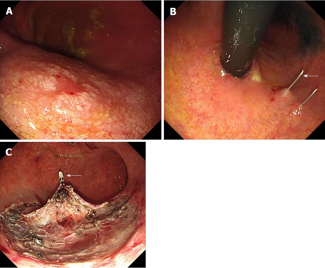
Figure 2 Results of gastric endoscopic submucosal dissection.
A: The remnant gastric lesion located at the middle posterior wall showed a mucosal cancer lesion about 10 mm in diameter; B: Just after the insertion of the scope into the stomach, Funada-type gastric wall fixation (arrow) (Create Medic, Tokyo, Japan) was performed at two opposite sites; C: A hemoclip (Olympus, Tokyo, Japan) with thread (arrow) was attached to the specimen, and the thread was pulled via the gastrostoma.
- Citation: Sasaki T, Uesato M, Ohta T, Murakami K, Nakano A, Matsubara H. Gastric endoscopic submucosal dissection via gastrostoma before the second operation for esophageal perforation: A case report. World J Gastrointest Endosc 2018; 10(6): 121-124
- URL: https://www.wjgnet.com/1948-5190/full/v10/i6/121.htm
- DOI: https://dx.doi.org/10.4253/wjge.v10.i6.121


