Copyright
©The Author(s) 2018.
World J Gastrointest Endosc. Nov 16, 2018; 10(11): 367-377
Published online Nov 16, 2018. doi: 10.4253/wjge.v10.i11.367
Published online Nov 16, 2018. doi: 10.4253/wjge.v10.i11.367
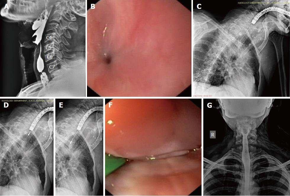
Figure 5 Near-total strictures in case 2 and their management.
A: Lateral view of the esophagogram showing the 2 near-total strictures; B: Endoscopic view through the right piriform sinus showing the tiny opening of the proximal near-total stricture. C: A flexible guide-wire was successfully passed across both the strictures; D, E: A 10F diathermic dilator being passed over the guide-wire to dilate the strictures. This was followed by balloon dilatation; F: Endoscopic view of electroincision (with a wire-guided sphincterotome) of the residual adhesions in the proximal stricture; G: The follow-up esophagogram at 2 wk of the completion of endoscopic therapy.
- Citation: Dhaliwal HS, Kumar N, Siddappa PK, Singh R, Sekhon JS, Masih J, Abraham J, Garg S. Tight near-total corrosive strictures of the proximal esophagus with concomitant involvement of the hypopharynx: Flexible endoscopic management using a novel technique. World J Gastrointest Endosc 2018; 10(11): 367-377
- URL: https://www.wjgnet.com/1948-5190/full/v10/i11/367.htm
- DOI: https://dx.doi.org/10.4253/wjge.v10.i11.367


