Copyright
©The Author(s) 2018.
World J Gastrointest Endosc. Nov 16, 2018; 10(11): 367-377
Published online Nov 16, 2018. doi: 10.4253/wjge.v10.i11.367
Published online Nov 16, 2018. doi: 10.4253/wjge.v10.i11.367
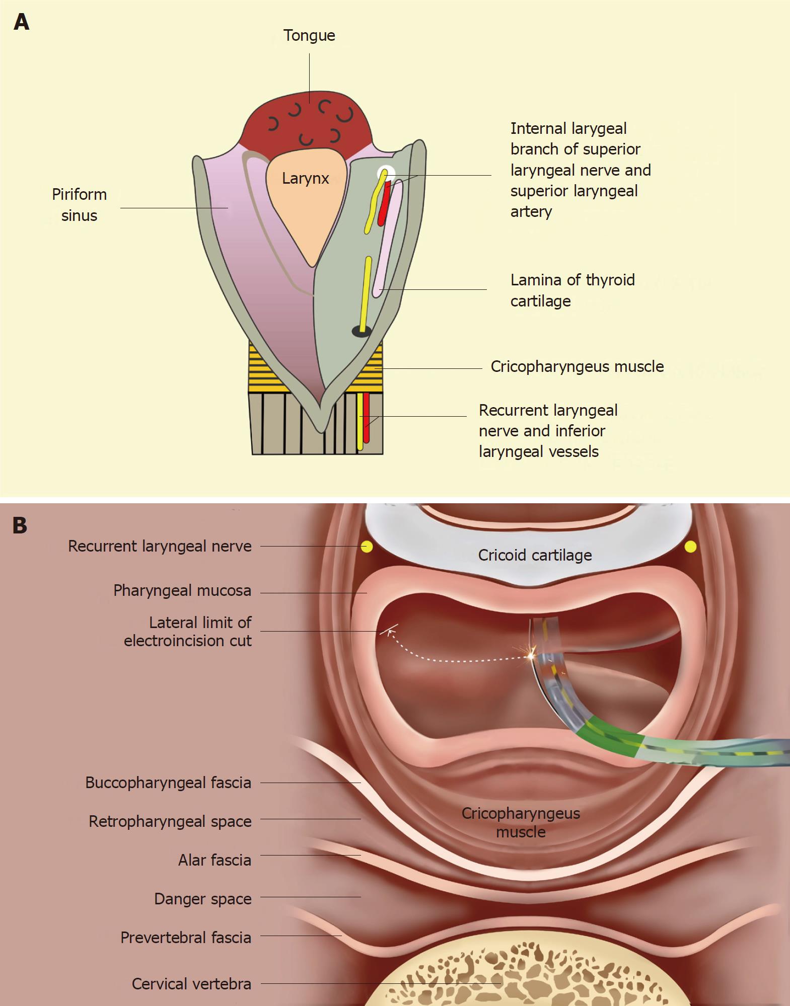
Figure 4 Anatomical considerations to prevent complications while performing the sphincterotome-assisted electroincision of the hypopharyngo-esophageal strictures.
A: An opened posterior view of the hypopharynx showing the submucosal location of the nerves and vessels within the anterior aspect of piriform sinuses. The internal branch of the superior laryngeal nerve lies cranially (proximally) while the recurrent laryngeal nerve lies caudally (inferiorly). An injury to the former may result in anesthesia of the laryngeal mucous membrane as far inferiorly as the vocal cords. An injury to the recurrent laryngeal nerve may lead to ipsilateral vocal cord paralysis. These neuro-vascular structures are important to be protected during the electroincision; B: Cross-sectional view at the level of cricopharynx showing the electro-incision of adhesions with a wire-guided sphincterotome. The sphinterotome is progressively moved laterally towards the pharyngeal wall (along the curved dashed line) with its wire cutting the adhesions in the post-cricoid space and then in the left piriform sinus. Care should be taken to stop few millimetres away from the anterior aspect of piriform sinus to avoid damage to the recurrent laryngeal nerve lying submucosally in this region. Similarly, the internal laryngeal nerve and vessel lie at the same location (in a more cranial cross-section) and need to be protected.
- Citation: Dhaliwal HS, Kumar N, Siddappa PK, Singh R, Sekhon JS, Masih J, Abraham J, Garg S. Tight near-total corrosive strictures of the proximal esophagus with concomitant involvement of the hypopharynx: Flexible endoscopic management using a novel technique. World J Gastrointest Endosc 2018; 10(11): 367-377
- URL: https://www.wjgnet.com/1948-5190/full/v10/i11/367.htm
- DOI: https://dx.doi.org/10.4253/wjge.v10.i11.367


