Copyright
©The Author(s) 2018.
World J Gastrointest Endosc. Nov 16, 2018; 10(11): 367-377
Published online Nov 16, 2018. doi: 10.4253/wjge.v10.i11.367
Published online Nov 16, 2018. doi: 10.4253/wjge.v10.i11.367
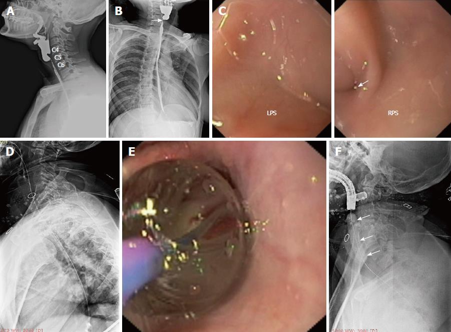
Figure 2 Near-total stricture in case 1 and its endoscopic management.
A: Lateral view of barium esophagogram showing the near-total stricture of the proximal esophagus involving the hypopharynx; B: On AP view, the near-total stricture has an eccentric proximal appearance (white arrow) and it is communicating with the right piriform sinus; C: During flexible endoscopic evaluation, the left piriform sinus (LPS) was completely obliterated in its apical region, while the right piriform sinus (RPS) showed a tiny opening (white arrow) which represents the beginning of the near-total stricture; D: A flexible guide-wire was passed across the stricture with difficulty, its correct placement was confirmed under fluoroscopy and the stricture was dilated with a 10F diathermy dilator (video 1); E, F: Subsequently, the stricture was dilated with a TTS balloon (white arrows).
- Citation: Dhaliwal HS, Kumar N, Siddappa PK, Singh R, Sekhon JS, Masih J, Abraham J, Garg S. Tight near-total corrosive strictures of the proximal esophagus with concomitant involvement of the hypopharynx: Flexible endoscopic management using a novel technique. World J Gastrointest Endosc 2018; 10(11): 367-377
- URL: https://www.wjgnet.com/1948-5190/full/v10/i11/367.htm
- DOI: https://dx.doi.org/10.4253/wjge.v10.i11.367


