Copyright
©The Author(s) 2018.
World J Gastrointest Endosc. Nov 16, 2018; 10(11): 367-377
Published online Nov 16, 2018. doi: 10.4253/wjge.v10.i11.367
Published online Nov 16, 2018. doi: 10.4253/wjge.v10.i11.367
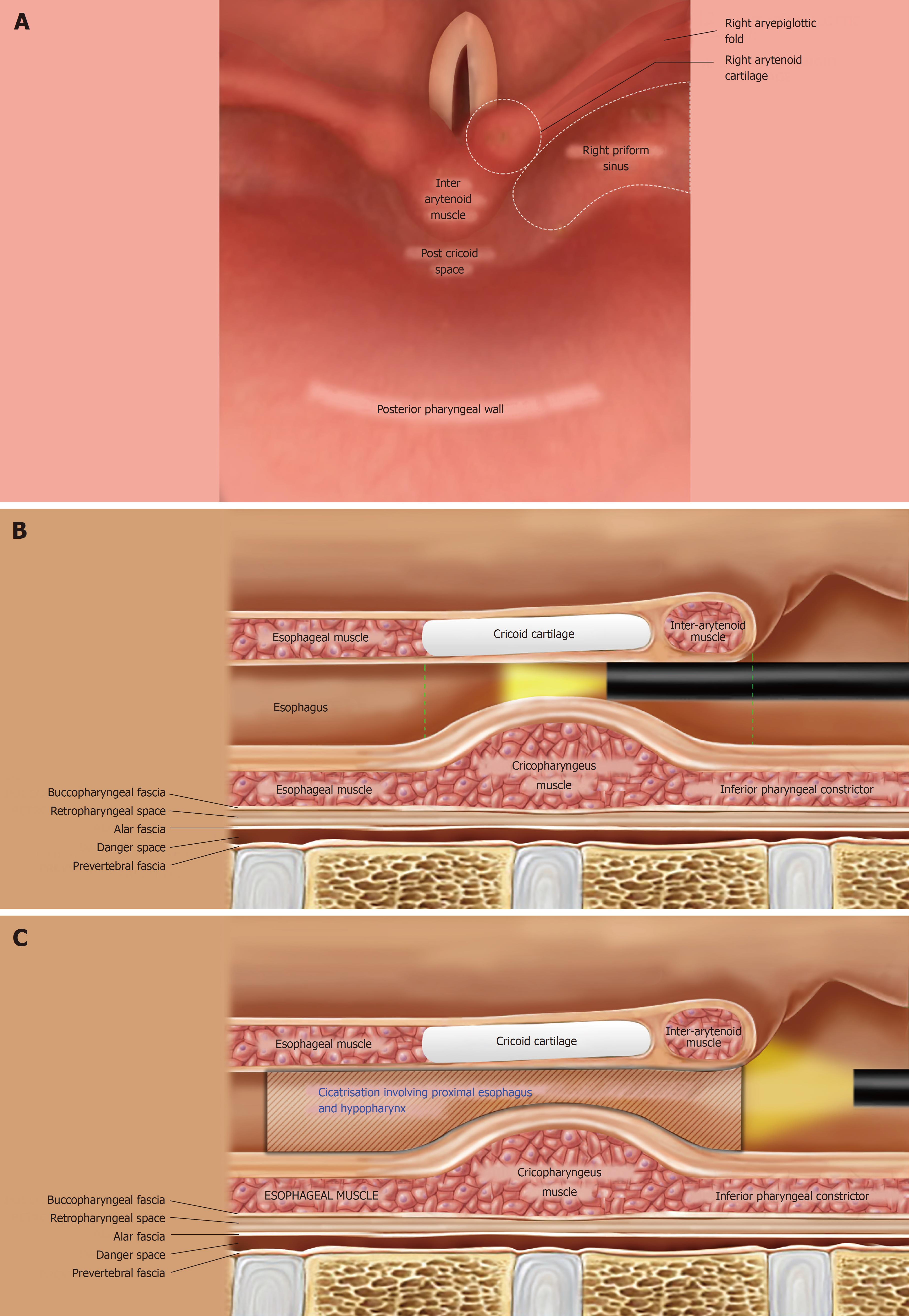
Figure 1 Anatomical consideration while addressing hypopharyngo-esophageal strictures endoscopically.
A: The normal anatomy of the adult hypopharynx as seen during flexible endoscopy, with the scope tip behind the epiglottis. The piriform sinuses are 2 inverted pyramids on either side of the larynx with their apex inferiorly at the level of cricopharyngeus muscle (CP); they are bounded medially by the aryepiglottic folds and laterally by the pharyngeal wall; B: Post-cricoid region extends from the level of arytenoid cartilages (joined by the inter-arytenoid muscle in the midline) superiorly to the inferior border of cricoid cartilage (vertical dashed green lines); C: The strictured proximal esophagus and hypopharynx in both of our cases. The fibrotic process in both extended cranially till the proximal aspect of the post-cricoid region; there was an involvement of the apical parts of piriform sinuses as well (not shown here).
- Citation: Dhaliwal HS, Kumar N, Siddappa PK, Singh R, Sekhon JS, Masih J, Abraham J, Garg S. Tight near-total corrosive strictures of the proximal esophagus with concomitant involvement of the hypopharynx: Flexible endoscopic management using a novel technique. World J Gastrointest Endosc 2018; 10(11): 367-377
- URL: https://www.wjgnet.com/1948-5190/full/v10/i11/367.htm
- DOI: https://dx.doi.org/10.4253/wjge.v10.i11.367


