Copyright
©The Author(s) 2018.
World J Gastrointest Endosc. Nov 16, 2018; 10(11): 326-339
Published online Nov 16, 2018. doi: 10.4253/wjge.v10.i11.326
Published online Nov 16, 2018. doi: 10.4253/wjge.v10.i11.326
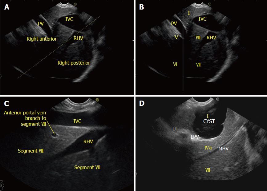
Figure 15 Liver sectors and liver segments are visualized.
A: The inferior vena cava (IVC) runs parallel to the probe in a long axis. A line along the right hepatic vein divides the liver into anterior and posterior sectors; B: The right lobe of the liver contains segments V to VIII. The segments are seen through the caudate process. The white line is drawn along the upper border of the curving part of the portal vein; C: The right hepatic vein is seen joining the IVC at an angle of around 60°. Segment VII is seen above the hepatic vein and segment VIII is seen between the hepatic vein and the IVC; D: The middle hepatic vein drains segment IV, segment V, and segment VIII. RHV: Right hepatic vein; LPV: Left branch of the portal vein; IVC: Inferior vena cava; PV: Portal vein.
- Citation: Sharma M, Somani P, Rameshbabu CS, Sunkara T, Rai P. Stepwise evaluation of liver sectors and liver segments by endoscopic ultrasound. World J Gastrointest Endosc 2018; 10(11): 326-339
- URL: https://www.wjgnet.com/1948-5190/full/v10/i11/326.htm
- DOI: https://dx.doi.org/10.4253/wjge.v10.i11.326


