Copyright
©The Author(s) 2018.
World J Gastrointest Endosc. Nov 16, 2018; 10(11): 326-339
Published online Nov 16, 2018. doi: 10.4253/wjge.v10.i11.326
Published online Nov 16, 2018. doi: 10.4253/wjge.v10.i11.326
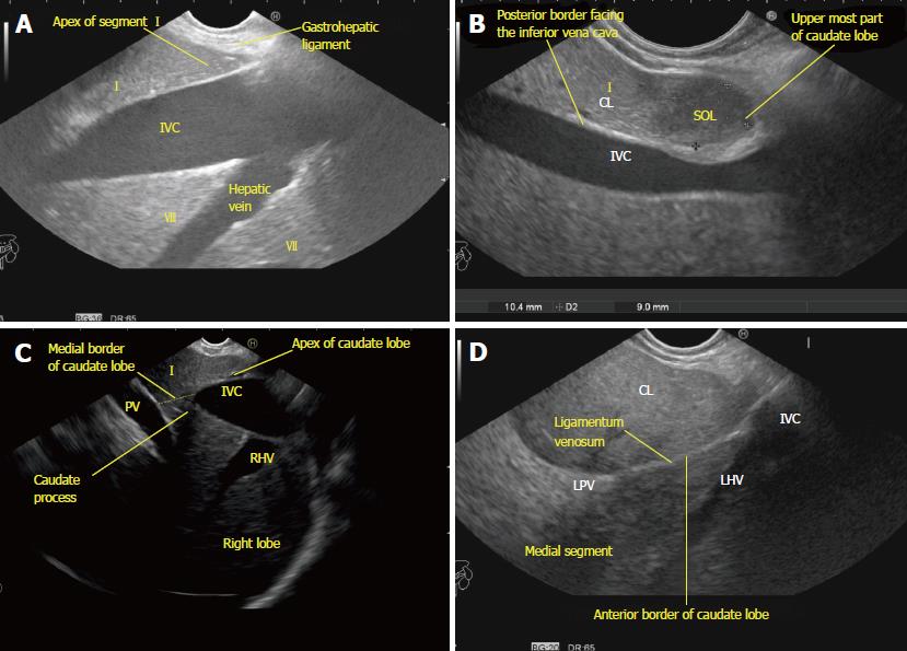
Figure 11 The caudate lobe, its boundaries, and its relationships.
A: The apex of the caudate lobe lies like a wedge near the joining of the hepatic veins. The base faces the inferior vena cava; B: A small metastatic space-occupying lesion is seen near the tip of the caudate lobe of the liver near the diaphragm between the probe and the inferior vena cava. Anterior margin of lesion is limited by the fissure for the ligamentum venosum; C: The continuity of the caudate lobe into the right lobe of the liver via the caudate process; D: The ligamentum venosum proceeds towards the umbilical part of the portal vein and divides the left medial segment from the caudate lobe. The ligamentum venosum is the anterior border of a pyramidal shaped caudate lobe. The attachment of the ligamentum venosum demarcates the lowest limit of the anterior border. SOL: Space-occupying lesion; LHV: Left hepatic vein; LPV: Left branch of the portal vein; RHV: Right hepatic vein; IVC: Inferior vena cava; PV: Portal vein.
- Citation: Sharma M, Somani P, Rameshbabu CS, Sunkara T, Rai P. Stepwise evaluation of liver sectors and liver segments by endoscopic ultrasound. World J Gastrointest Endosc 2018; 10(11): 326-339
- URL: https://www.wjgnet.com/1948-5190/full/v10/i11/326.htm
- DOI: https://dx.doi.org/10.4253/wjge.v10.i11.326


