Copyright
©The Author(s) 2018.
World J Gastrointest Endosc. Nov 16, 2018; 10(11): 326-339
Published online Nov 16, 2018. doi: 10.4253/wjge.v10.i11.326
Published online Nov 16, 2018. doi: 10.4253/wjge.v10.i11.326
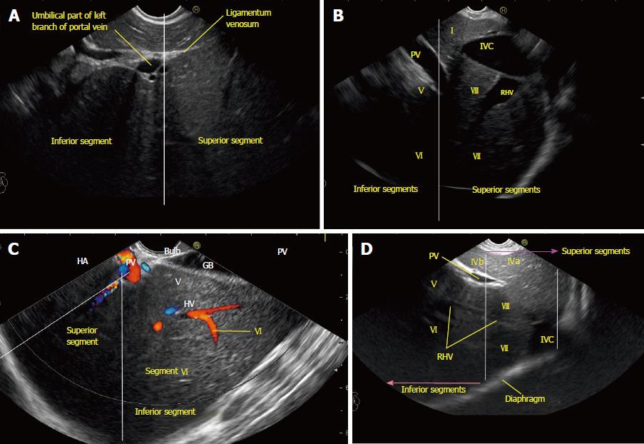
Figure 10 The division of superior and inferior liver segments by the portal vein and its branches.
A: Line at the level of the upper border of the umbilical segment of the portal vein dividing the superior and inferior segments; B: The white line divides the superior and inferior segments. This diagram from the esophagus shows the right side of the liver through the caudate lobe of the liver and the inferior vena cava. The presence of hepatoduodenal ligament around the portal vein may not allow a similar quality of visualization of the inferior segments (V and VI); C: This image from station 2 (duodenum bulb) shows the right and middle hepatic veins. The right hepatic vein is parallel to the surface of the gallbladder, and the middle hepatic vein is towards the neck of the gallbladder. Only the right lobe is seen through the gallbladder; D: The right hepatic vein drains segments VI and VII and a variable portion of segments V and VIII. Segment I has direct drainage into the intrahepatic/retrohepatic part of the inferior vena cava. RHV: Right hepatic vein; IVC: Inferior vena cava; PV: Portal vein.
- Citation: Sharma M, Somani P, Rameshbabu CS, Sunkara T, Rai P. Stepwise evaluation of liver sectors and liver segments by endoscopic ultrasound. World J Gastrointest Endosc 2018; 10(11): 326-339
- URL: https://www.wjgnet.com/1948-5190/full/v10/i11/326.htm
- DOI: https://dx.doi.org/10.4253/wjge.v10.i11.326


